Transcription of THORACIC PARAVERTEBRAL BLOCK: LANDMARK …
1 Sign up to receive ATOTW weekly - email ATOTW 224 PARAVERTEBRAL block , LANDMARK Techniques 23/05/2011 Page 1 of 10 THORACIC PARAVERTEBRAL block : LANDMARK TECHNIQUES ANAESTHESIA TUTORIAL OF THE WEEK 224 23RD MAY 2011 Dr Mark Dodd and Dr John Hunsley Northern General Hospital, Sheffield, UK Correspondence to QUESTIONS Before continuing, try to answer the following questions. The answers can be found at the end of the article, together with an explanation. 1. Which one of the following statements is incorrect? a. The PARAVERTEBRAL space communicates laterally with the intercostal space and medially with the intervertebral foramen. b. When performing a THORACIC PARAVERTEBRAL block sympathetic blockade is essential for reliable postoperative analgesia. c. In a 70kg patient 5 dermatomes can safely be anaesthetised using 5 injections, each of 5ml bupivacaine. d. The superior aspect of the tip of the spine of T2 lies adjacent to the transverse process of T2. 2. Which of the following are relative contraindications to THORACIC PARAVERTEBRAL block ?
2 A. Ipsilateral empyema b. Mesothelioma c. Competent adults declining the procedure d. Previous anaphylaxis to procaine. 3. Before surgery, under general anaesthetic and 20 minutes after insertion of a PARAVERTEBRAL block a patient has a cardiac arrest. Which of the following are possible causes? a. Local anaesthetic toxicity b. Hypotension due to sympathetic blockade c. Anaphylaxis d. Tension pneumothorax INTRODUCTION In 1905 Hugo Sellheim performed the first documented THORACIC PARAVERTEBRAL block (TPVB) in Leipzig. Originally the technique was used to provide muscle relaxation and anaesthesia for upper abdominal The introduction of curare and volatile gases replaced the need for TPVB. Concerns about the safety of the block accentuated the decline of its use. In 1979 a seminal paper describing a loss of resistance technique reignited interest in Evidence and experience in the use of PARAVERTEBRAL blockade has mounted. It can be used for abdominal, THORACIC and breast surgery, chronic pain and other niche areas (see indications below).
3 The safety concerns from previous decades have proved to be unfounded with TPVB having a comparable rate of complications to THORACIC epidurals and intercostal As TPVB use has risen other possible clinical benefits have been explored. Early research has suggested a reduction in breast cancer recurrence in patients where TPVB was used for their initial Sign up to receive ATOTW weekly - email ATOTW 224 PARAVERTEBRAL block , LANDMARK Techniques 23/05/2011 Page 2 of 10 A large phase III multinational, multicenter trial is presently recruiting to examine breast cancer five year recurrence rates in patients receiving TPVB compared to morphine ANATOMY PARAVERTEBRAL blockade (PVB) is achieved by placement of local anaesthetic around the nerve bundles as they arise from their corresponding intervertebral foramina. PVB is often used without prefix to refer to THORACIC PVB (TPVB). It should be noted that cervical stellate ganglion block ), lumbar PARAVERTEBRAL blocks and lumbar plexus blocks are also types of PVB.
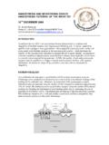
4 For clarity a spinal level should be stated when describing the block as the anatomy will obviously differ. The THORACIC PARAVERTEBRAL space (TPVS) is a wedged shape with the apex lying laterally and the base medially as shown in Figure 1. Figure 1: Drawing of the THORACIC PARAVERTEBRAL space. The boundary of the space is depicted by a transparent wedge. Relationships Anterolaterally (from posterior to anterior) lie the parietal pleura, the pleural space, visceral pleura and lung parenchyma. Medially lies the posterolateral portion of the vertebral body, the vertebral disc and the vertebral foreman with its corresponding spinal nerve. Posteriorly the TPVS is limited by the superior costotransverse ligament. Laterally the space is bound by the posterior intercostal membrane and the intercostal space. Sign up to receive ATOTW weekly - email ATOTW 224 PARAVERTEBRAL block , LANDMARK Techniques 23/05/2011 Page 3 of 10 Communications The TPVS is continuous from T1 to T12.
5 For descriptive purposes the space is split into dermatomes and each segment of the PVS is said to be limited superiorly and inferiorly by the heads of the corresponding ribs. The cervical and THORACIC PVS are directly continuous with one another. There are conflicting contrast studies in cadavers with regards a communication between the THORACIC and lumbar PVS. Clinically lumbar plexus block is rarely seen following lower TPVB. The TPVS communicates medially with the epidural space via the intervertebral foramina and laterally with the intercostal spaces. The prevertebral fascia lies anterior to the vertebral bodies and can provide a conduit to the contralateral TPVS for local anaesthetic but this is unusual. The TPVS may be divided into anterior and posterior segments by the endothoracic fascia. This a thin fibroelastic structure and may affect the pattern of spread of local anaesthetic during TPVB. The existence of the endothoracic fascia is debated.
6 Nerves The nerve root passes through its respective intervertebral foramen to enter the medial aspect of the TPVS. There is no fascial sheath covering the nerve as it emerges as a loose bundle of neurones. This allows for direct and quick action of local anaesthetic on the neurones. Each root projects a somatic dorsal ramus and a ramus communicantes within the medial aspect of the TPVS. The larger ventral portion passes through loose areolar tissue and exits the TPVS via the corresponding intercostal space. The sympathetic chain lies on the neck of the ribs lateral to the heads and anterior to the intercostal bundles. Nociceptive pathways are diverse and multiple and include fibres passing through the sympathetic chain. Blockade of the chain is therefore essential for effective analgesia. INDICATIONS TPVB lends itself to unilateral surgery but bilateral TPVB have been described. More superficial surgery such as inguinal hernia repair, mastectomy and breast augmentation can be performed using PARAVERTEBRAL block as the sole anaesthetic technique.
7 Table 1: Clinical applications of THORACIC PARAVERTEBRAL blocks. THORACIC SURGERY TPVB will provide analgesia for chest wall incisions, drain sites and the parietal pleura. Open thoracotomy Video assisted thoracoscopic surgery (VATS) Breast surgery including axillary dissection Minimally invasive cardiac surgery ABDOMINAL SURGERY Incisions around the midline will require bilateral TPVB and lower abdominal incisions often require additional upper lumbar PVB. Renal surgery Open and laparoscopic Cholecystectomy Appendicectomy Inguinal Hernia repair NON-SURGICAL MANAGEMENT Postherpetic neuralgia Post thoracotomy chronic pain Fractured ribs Maternal Labour Angina pectoris Pain from mesothelioma SYMPATHETIC BLOCKADE Injection of local anaesthetic into the TPVS will result in localised sympathetic block resulting in a fall of blood pressure. This is usually less marked than that seen following THORACIC epidural. TPVB are rarely used for sympathetic blockade. Therapeutic control of hyperhydrosis SVT Sign up to receive ATOTW weekly - email ATOTW 224 PARAVERTEBRAL block , LANDMARK Techniques 23/05/2011 Page 4 of 10 CONTRAINDICATIONS Before any regional procedure a risk assessment must be made and balanced against the patients requirements.
8 Perhaps the only absolute contraindications are patients who have an allergy to local anaesthetic or whom decline the procedure. Contraindications should be considered in the context of the surgery, the patient, co-morbidity and alternative forms of analgesia available. Table 2: Considerations before insertion of a TPVB. CONSIDERATION CONCERN Patient refusal Absolute contraindication Anticoagulation / clotting abnormality The PVS is not easily accessible to stem bleeding. It is an expandable space of significant size should bleeding be caused by needle trauma. A similar approach as for epidurals should be taken. Local infection cellulitis, abscess) Concerns of seeding infection. Previous ipsilateral THORACIC surgery Fibrosis and scaring may destroy or distort the PARAVERTEBRAL space and make pleural puncture more likely. A loss of resistance technique may be ineffective in the presence of scarring. Spread on local anaesthetic may be more unpredictable Total pleurectomy TPVB relies upon local anaesthetic remaining within the TPVS to act upon the nerves within it.
9 The parietal pleura forms the anterior border of the space. If it is absent local anaesthetic within the TPVS will spread away between the lung surface and the chest wall. Localised tumour The use of TPVB for thoracotomy is often within the context of lung, pleural or breast malignancy. Chest wall or TPVS involvement may prevent the use of TPVB due to concerns of seeding malignant cells or bleeding. If a malignant effusion is tapped the needle should be removed without injecting the mixture of local anaesthetic and pleural fluid. Empyema A successful TPVB does not breach the parietal pleura but consideration should be given to accidental puncture in the presence of an empyema. Presence of a catheter in proximity to infection should be avoided. Abnormal THORACIC anatomy The anteroposterior distance of the TPVS is narrow. This may be altered with THORACIC kyphosis or scoliosis. Mass effect of effusions and tumour may also distort the anatomy.
10 Previous spinal surgery may cause scarring and disruption of the superior costotransvere ligament making loss of resistance techniques unreliable and ultrasound guidance difficult. TECHNIQUES As with all regional anaesthetic procedures: consent must be obtained from the patient; iv access established and standard monitoring attached; resuscitation facilities must be available and the procedure carried out in an aseptic manner. Spread The TPVB has been shown to have an unpredictable spread. A large volume injected at a single space may be confined to that space, spread longitudinally across several dermatomes or both. The pattern of spread may depend upon whether the anaesthetic is injected into the dorsal or ventral compartment of the TPVS. The endothoracic fascia, if present, is thin and not amenable to identification by loss of resistance or ultrasound. Sign up to receive ATOTW weekly - email ATOTW 224 PARAVERTEBRAL block , LANDMARK Techniques 23/05/2011 Page 5 of 10 A large volume, single injection technique will provide analgesia/anaesthesia for the dermatome level at which it is injected.