Transcription of Tibial Plateau Fractures - Orthopaedic Trauma Association ...
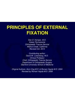
1 Tibial Plateau Fractures :Evaluation, Assessment and TreatmentThuan V. Ly, MDOhio State UniversityObjectives Recognize the anatomy of the proximal tibia Describe initial evaluation and management Identify common fracture patterns Apply treatment principles and strategies Partial articular Fractures Complete articular Fractures Discuss rehabilitation , complications, and outcomes Illustrate selected Tibial Plateau casesEpidemiology (burden of disease/cost to society) Tibial Plateau Articular surface proximal tibia +/-metaphyseal /diaphyseal extension Account for of all Fractures Lateral Plateau : 55-70% of Fractures Medial Plateau : 10-20% of Fractures Bicondylar Plateau : 10-30% of fracturesEpidemiology(burden of disease/cost to society) Bimodal distribution Young adults: high energy mechanism Highest in 5thdecade Male > Female Elderly: low energy mechanism Osteoporotic bone Female > Male Significant functional impairment Joint incongruity, malalignment, instability Post-traumatic arthritisAnatomy Consist of medial and lateral Plateau Medial larger Medial lower (concave) Medial bone harder (thus less likely to fracture) Lateral higher (convex) Lateral cartilage thicker (3 4 mm)MedialLateralAnatomyMedialconcaveLate ralconvexAnatomy Bony prominences Intercondylar eminence (menisci & cruciate ligaments attachment) Tibial tubercle (patellar tendon) Gerdy s tubercle (Iliotibial band) Tibia slope.
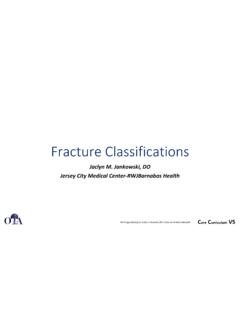
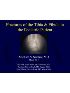
2 10 degrees posteroinferiorAnatomy Lateral meniscus Larger (cover more articular surface) Commonly torn with lateral Plateau fracture Medial meniscus C shapedMedialLateralMechanism of Injury Valgus producing force Lateral Plateau Varus producing force Medial Plateau Axial compressive force Bicondylar Plateau Combination High energy Bicondylar Plateau Soft tissue injuryMechanism of Injury Valgus producing force Lateral Plateau Varus producing force Medial Plateau Axial compressive force Bicondylar Plateau Combination High energy Bicondylar Plateau Soft tissue injuryMechanism of Injury Valgus producing force Lateral Plateau Varus producing force Medial Plateau Axial compressive force Bicondylar Plateau Combination High energy Bicondylar plateauMechanism of Injury Low energy Split depression Increasing age Poor bone quality High energy Pedestrian car (bumper)
3 Fall from height Motor vehicle accident Axial load (knee extended) Bicondylar fracture Associated injuriesAssociated Injuries Ligaments MCL, LCL ACL, PCL Menisci Lateral meniscus likely if: > 5mm depression > 6mm condylar widening Gardner J Trauma 2006 Popliteal artery Peroneal nerve Compartment syndromeMCL tearAssociated Injuries Lateral Plateau Tear of meniscus MCL / ACL tear Medial Plateau Fracture / dislocation variant Popliteal artery injury Peroneal nerve injury Bicondylar Open injury Compartment syndromeEvaluation -History Mechanism of injury Injury factors Soft tissues Fracture patterns Associated injuries Patient factors Age Bone quality Comorbidities Previous level of activity Function demandsEvaluation Physical Exam Initial Inspection Skin integrity Soft tissue swelling Open fracture Gross deformity Shortened limb Neurovascular status Document the Exam!
4 !!Evaluation Physical Exam Low energy mechanism Knee swelling Limited knee ROM Tender to palpation Able to assess knee stability Varus/valgus stress 0 and 30 degrees Lachman s exam for ACL deficiencyEvaluation Physical Exam High energy mechanism Advanced Trauma Life Support (ATLS) Resuscitation Limb threatened Soft tissue integrity Open fracture Abrasions Blisters Compartment syndrome Knee stability exam Difficult to performEvaluation Physical Exam Soft tissue assessment Know Gustilo & Anderson open Fractures classification Tscherne -closed Fractures classification Avoid missing compartment syndrome Determine timing of surgery Skin wrinkles present?Evaluation Physical Exam Document NV status Neurologic Peroneal nerve Vascular Ankle-Brachial Index ABI > by AO Foundation, SwitzerlandEvaluation Physical Exam ABI Screening test LE injuries with concerns for vascular injury Obtain systolic pressure Uninjured upper extremity (Brachial) Injured LE limb (Ankle)
5 BP cuff just proximal to the ankle DP or PT pulseEvaluation Physical Exam ABI < Predictable of arterial injury Vascular consult Proceed with arteriogram ABI > Admit for observation Followed with serial noninvasive exam Johansen et al J Trauma Injured Extremities ABI Sensitivity = 95% Specificity = 97% Mills et al J Trauma 2004 Knee dislocation ABI Sensitivity and Specificity = 100%Evaluation -Radiographic Plain X-ray knee/tibia AP Lateral Obliques of knee Internal or external rotationEvaluation -Radiographic Tibial Plateau view Normal Tibial slope 10 degrees posteroinferior10 degreesAngle for x-rayEvaluation -Radiographic CT scan Surgical consideration exists Complex Fractures to assist in surgical planning Assessing Depression Comminution Fracture line (coronal split-medial side with bicondylar Plateau ) Obtain CT afterapplying traction (ex fix)Evaluation -Radiographic MRI scan?
6 Subtle nondisplaced fracture line Gardner JOT 2005 Noted high associated soft tissue injuries Lat. meniscus : 91% Med. meniscus 44% ACL PCLC lassification Schatzker Type I: Split fracture of the lateral Plateau Type II: Split depression fracture of the lateral Plateau Type III: Pure depression fracture of the lateral Plateau Type IV: Medial Plateau (possible fracture / dislocation) Type V: Bicondylar Plateau fracture Type VI: Plateau fracture with metaphyseal / diaphyseal dissociationClassification AO / OTA (41-Proximal section) Type A: Extraarticular fracture (41-A) Type B: Partial articular fracture (41-B) B1: Pure split B2: Pure depression B3: Split depression Type C: Complete Articular fracture (41-C) C1: Simple articular, Simple metaphyseal C2: Simple articular, Multi-fragmentary metaphyseal C3: Multifragmentary articularClassification Unicondylarfracture SchatzkerI, II, III AO/OTA (41-B) Partial articularSplitSplit-depressionCentral depressionClassification Unicondylar fracture Schatzker IV AO/OTA (41-B) Partial articular Medial Plateau Fracture / dislocation Displaced, higher energy Vascular injury concernSplit fracture, medial plateauClassification Bicondylar fracture Schatzker V, VI V: Medial Tibial Plateau split and Lateral split depression VI: Plateau with metadiaphyseal dissociation AO/OTA (41-C) Complete articularBicondylar fractureMetadiaphyseal dissociationTreatment Principles Soft tissue management Surgical timing is important Wringles in the skin Temporary Stabilization Staged protocol Barei et al.
7 JOT 2004 Egol et al. JOT 2005 Treatment Principles Anatomic reduction of articular surface Obtain and maintain Reduce condylar width Address meniscal injuries Restore mechanical axis metadiaphysis Stable fixation Early ROMT reatment Options: Nonsurgical Patient factors Elderly Nonambulatory Pre-existing arthritis Injury factors Articular incongruity <5 mm, elderly, sedentary activity Stable Varus / Valgus stress < 5 -10 degrees instability71 y/o male, multiple med. comorbiditiesNonsurgical Technical Pearls Immobilize 1-2 weeks Knee immobilizer or hinge knee brace Locked in extension Start ROM Controlled motion Start 0-30 degrees and advance as tolerated Goal-90 degrees at 4wks NWB 6-8 weeksRadiographic F/UWeekly for first 3 weeksIndications for Surgery Absolute indications Open Tibial Plateau Associated compartment syndrome Associated vascular injuryIndications for Surgery Relative indications Axial malalignment Instability in full extension Articular incongruity >3mm in young, active Condyle wideningIndications for Surgery Displaced bicondylar Most if not all medial plateauTiming of SurgeryLow Energy: Fixed electively and earlyHigh Energy.
8 Be patienceTemporary External Fixation Knee spanning external fixation Ligamentotaxis Improve fracture fragment gross alignment Length and alignment Minimize further damage to articular surface Soft tissue assessment and wound careTemporary External Fixation Candidates for external fixation Axially unstable Tibial Plateau fracture Bicondylar fracture Schatzker type V and VI Fracture / Dislocation Schatzker type IVExternal Fixation: Patient set up Supine Radiolucent operating table C-arm fluoroscope Contralateral side Sterile towel bump Allow 5-10 degrees knee flexion 2 pins in femur Anterior or lateral 2 pins in tibia Antero-medialImplants External Fixation Large external fixator system 5 mm half threaded schanz pins Self drilling Different length available Connecting rods and Clamps Compressive dressing Ext. fix sponges Retention clipExternal Fixation -Pearls Mark knee joint and fracture sites Schanz pins placement out of zone of future surgical incisions Pre-drilling for good bone quality Avoid skin tension by pins Pin spread to improve construct stabilityExternal Fixation -Pearls Placement of metal clamps Away from knee joint and fracture zone Allows better imaging Padded prefabricated posterior splint Offload heel Compressive dressing Stabilize pin-skin interface Minimize pin-skin motionTemporary Stabilization-Case Example Staged protocol Knee spanning external fixation Restore length, alignment, rotation Definitive ORIF 10-21 days Wait for soft tissue CT scan Preop planORIF-Patient Set Up Radiolucent operating table C-arm fluoroscope Contralateral side of injured limp Exception.
9 Medial Plateau -ipsilateral side Buttock bump Tourniquet Extremity positioners Sterile towel bump Leg ramp Radiolucent for imagingPatient Set Up-Technical Pearls IV bag pump-buttock bump Deflated allows easier access to posteromedial tibiaPatient Set Up-Technical Pearls IV bag pump-buttock bump Inflated allows neutral leg alignment for anterolateral approachORIF-Equipment Headlamps Femoral distractor Osteotomes Bone tamps Fracture reduction instruments K-wiresORIF-Implant options Unicondylar fracture Conventional non-locking plate L or T plate Buttress Pre-contoured periarticular plates Raft screws alone or Locking plate Osteoporotic boneORIF-Implant Options Angular stable (Locking) implants Precontour for proximal tibia Bicondylar tibia Plateau with metadiaphyseal involvement Spanning or bridging across fracture zone Selected fracture, allows stabilization of medial plateauExternal Fixation Limited internal fixation Small incisions or percutaneous Thin-wire ring fixators Connect to the shaft Fixation distally with 5mm half-pins Advantages Minimize soft tissue injury Still need to reduce articular surface!
10 !!ORIF-Fixation Summary Fixation based on fracture type Type I, II, III: Buttress plates with raft screws Type IV: Medial plate (buttress) Be cognizant of any impaction of lateral joint line Type V, VI: Important to understand plate function Pattern dictates fixation Single lateral base fixed angle implant Dual plating (lateral and posteromedial) Surgical Approaches Anterolateral Lateral Plateau involvement Combination with medial for complex Plateau Posteromedial Medial Plateau Coronal split Posterior Dual approaches Anterolateral PosteromedialCopyright by AO Foundation, SwitzerlandSurgical Approach: Anterolateral Most common approach Lazy S or Inverted L Curvilinear incision centered over Gerdy s tubercle Extend distally of the anterior compartment fascia 1 cm off Tibial crest Subperiosteal elevate muscle Extend proximally midaxial line of knee joint Full thickness skin flapsSurgical Approaches: Anterolateral Incise and elevate IT band and anterior compartment fascia Subperiosteal dissection off lateral Tibial crest and not thru compartment muscle Submeniscal arthrotomy Inspect the meniscus Tag repair as neededSurgical Approach: Posteromedial Straight incision Posterior border of proximal tibia Avoid Saphenous nerve and vein Interval between Medial head gastrocnemius and hamstrings (Pes anserine tendons)Copyright by AO Foundation, SwitzerlandSurgical Approach.