Transcription of Trauma: Spinal Cord Injuries - - RN.org®
1 trauma : Spinal cord Injuries Reviewed January, 2017, Expires January, 2019 Provider Information and Specifics available on our Website Unauthorized Distribution Prohibited 2017 , , , LLC By Wanda Lockwood, RN, BA, MA The purpose of this course is to explain different types of traumatic Spinal cord Injuries , including primary and secondary Injuries , assessment, and management. Upon completion of this course, the healthcare provider should be able to: Describe the anatomy of the Spinal cord . Describe the 4 sections of the vertebral column. List the differences between upper motor neuron and lower motor neuron damage. Describe the functions related to different levels of Spinal nerves.
2 Describe the ABCDEs of initial assessment. Discuss secondary assessment. Describe the Glasgow Comas Scale, Describe 3 criteria for classification of Spinal cord Injuries . Describe sensory, manual muscle, and reflex testing. Describe the ASIA Impairment Scale (AIS). Describe the Canadian C-spine rule. Describe the pathophysiology of Spinal cord injury. Differentiate among 3 types of shock. Discuss respiratory complications. Discuss the use of steroids and traction. Discuss management of DVT, PE, urinary retention, thermoregulation, pressure sores, GI problems, metabolic abnormalities, and autonomic dysreflexia. Differentiate between quadriplegia and paraplegia.
3 Describe 6 cord syndromes. Purpose Goals Introduction Spinal cord Injuries have resulted in paralysis of over million people in the United States with about 10,000 new Injuries each year. The reasons vary, but work Injuries (28%), motor vehicle accidents (24%) and sporting accidents (16%), primarily diving, cause the most Injuries . In many cases, people suffer from multiple traumas and may, for example, also have brain injury. Young males are the most at risk for Spinal cord Injuries , and gunshot wounds are an increasing cause of injury. Approximately half of all Spinal cord Injuries involve the cervical spine (primarily C4 to C7), and half of Spinal cord Injuries result in complete quadriplegia.
4 The Spinal cord extends as a continuous structure from the medulla at the base of the skull to the first lumbar vertebra (L1), where it tapers into a fibrous band called the conus medullaris. At L2 the nerve roots (cauda equina) extend beyond the conus. The Spinal cord is approximately 18 inches (45 cm) long in an adult and about finger width. The vertebral column comprises 7 cervical, 12 thoracic, 5 lumbar, and 5 fused sacral vertebrae that protect the Spinal cord . Intervertebral discs and facet joints cushion and allow movement. Nerve roots exit from the vertebral column through the intervertebral foramina (openings). In the Spinal cord , gray matter is at the center in an H-shape and is surrounded by white matter that contains both afferent (ascending) and efferent (descending) nerve fibers.
5 Like the brain, the Spinal cord is surrounded by the meninges. The Spinal cord contains 31 pairs of Spinal nerves: 8 cervical, 12 thoracic, 5 lumbar, 5 sacral, and 1 coccygeal. Each nerve has a dorsal root and a ventral root. The dorsal roots transmit sensory information, such as temperature, proprioception, touch, and pain, from specific areas of the body, known as dermatomes. The ventral roots transmit motor impulses. There are 6 ascending tracts: 2 conduct sensation (touch, vibration, position, pressure), 2 conduct sensory impulses necessary for coordinated muscle contraction, and 2 conduct sensation of pain, temperature, fine touch, and vibratory sense from the upper body. There are 8 descending tracts (upper motor neurons): 2 conduct muscle impulses and control voluntary muscle activity, 3 involve autonomic functions (perspiration, circulation, pupil dilation) and involuntary muscle control, 1 conducts impulses for voluntary head and facial muscle movement, and the last 2 involve voluntary muscle movement.
6 Some of the motor nerve pathways, contained in the Spinal cord , represent the pathways of the extrapyramidal system (making connections from the anterior horn cells to the automatic control centers in the brain) and others are components of reflex arcs. Spinal cord injury can result in lesions of upper motor neurons and/or lower motor neurons. Upper motor neuron damage Lower motor neuron damage Loss of voluntary control (paralysis). Increased muscle tone. Muscle spasticity. No muscle atrophy. Hyperreflexia. Loss of voluntary control (paralysis). Decreased muscle tone. Muscle flaccidity. Muscle atrophy. Hyporeflexia. The nerves at different levels of the Spinal column control various functions, so Injuries result in predictable outcomes.
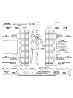
7 Level Functions C1-C6 Neck flexors. C1-T1 Neck extensors. C3, C4, C5 Innervate diaphragm. C5, C6 Shoulder movement. Raises arm (deltoid). Flexes elbow (biceps). C6 Supinates arm. C6, C7 Extends elbow (triceps), wrist (extensors). Pronates wrist. C7, T1 Flexes wrist. Innervates small muscles of hand. T1-T6 Innervates intercostals and upper trunk (above waist). T7-L1 Innervates abdominal muscles. L1-L4 Thigh flexion L2-S1 Thigh abduction L5-S2 Leg extension at hip (gluteus maximus). Plantar flexion of foot. Toe flexion. L2-L4 Leg extension at the knee (quadriceps femoris). L4-S2 Leg flexion at the knee (hamstrings). L4-S1 Toe extension. S2-S4 Bladder and bowel function/control. Initial assessment/Intervention With suspected Spinal cord or vertebral injury, the patient should be immediately immobilized as an estimated 3 to 25% of Injuries to the Spinal cord occur during transport or resuscitation.
8 All patients with pain along the spine or paresis/paralysis should be assumed to have Spinal cord Injuries until appropriate evaluation can be completed. The standard ABCDE evaluation should be completed as well as cranial nerve assessment, followed by a more extensive neurological examination once the patient has stabilized. Because Spinal cord Injuries are often associated with other types of Injuries , such as traumatic brain and/or abdominal Injuries and fractures, the evaluation must focus on identifying all possible Injuries . Airway Examine the airway for obstructions, such as loose teeth, foreign bodies. Lacerations and bone instability may be obstructive. Examine the trachea for deviation and observe for signs of circumoral cyanosis (sign of hypoxia).
9 Auscultate the airway and listen for turbulence. With Spinal cord injury, prevertebral swelling and hematoma may occur, and this can compromise the airway. Breathing Immediate intubation may be indicated for high cervical Injuries , but care must be used to avoid flexion of the neck. Manual inline immobilization or fiberoptic intubation is recommended. Neurologic status must be assessed along with pulmonary and respiratory function. High diaphragmatic/abdominal breathing is an indication of high cervical injury. Circulation Monitor blood pressure, pulse, temperature, color, and indications of cyanosis (circumoral, peripheral), including oxygen saturation continuously. Use venous access to restore intravascular volume, BP, and perfusion.
10 Primary assessment Evaluate possible causes for hypotension, a common finding with SCI, often indicating bleeding from other Injuries . Hypotension found with bradycardia often indicates Spinal cord injury. Note skin temperature. Warm skin may indicate adequate perfusion or neurogenic shock. Dysfunction/ disability Assess responsiveness Glasgow Coma Scale (GCS). Assess neurological status. Assess motor ability by observation, pressure to nail bed, or sternal rub: o Decreased spontaneous movement and/or flaccidity may be associated with local injury or Spinal cord injury. Assess reflexes to determine level of injury and integrity of the Spinal cord . Immobilize patient with rigid backboard and cervical spine collar until Spinal cord injury is ruled out.