Transcription of Ultrasound Imaging System - University of California, Los ...
1 Ultrasound Imaging System George Saddik, University of California, Los Angeles, CA Bioengineering Department Feb. 9th 2015 Today s Topics History What is Ultrasound ? Physics of Ultrasound Ultrasonic echo Imaging Focusing technique A-mode signal and B-mode image Features of echo image Transducers Design and Modeling Applications and Transducers History 1877 Discovery of piezoelectricity (Pierre and Jacques Curie) 1913 First sonar patent filed after Titanic disaster 1917 Sonar used for detecting range of u-boats during WWI Developed by the French government (Langevin student of Curies) Hydrophone hung over side of ship 1929-1935 Use of Ultrasound waves in detecting flaws in metals (Sokolov -USSR)
2 First patent for using Ultrasound waves to detect flaws in solids (Mulhauser, 1931) 1930s Ultrasound used for physical therapy for Europe s football teams Ultrasound used for sterilization of vaccines and for cancer therapy 1940s Ultrasound was seen as a cure-all therapy tool Used for arthritis, gastric ulcers, and eczema History Post WWII Surplus Naval sonar equipment used for medical applications Based on radar and sonar techniques Japanese led the development of medical sonography in 1940s Pulse-echo measurements on oscilloscopes Detection of gallstones, breast masses, and tumors Austrian group generated images of brain tumors and cerebral ventricles through skull Researchers from US Naval Medical Research Institute imaged gallstones 1950s Simple 2D Imaging devices developed by researcher in US and Japan Some were as large as a room The profession sonographer was created by the AMA Echocardiography (Sweden) Doppler measurements of tissue motion and blood flow (Japan, US) Focused Ultrasound ablation (Fry, U.)
3 Illinois Urbana) History 1970s Sonic Boom Medical sonography became accepted for several clinical applications Sector scanners, arrays Static 2D grayscale images, then real-time images CW and PW Doppler Fetal heart monitoring Dept. Education defined curriculum for sonographers Growth of NDT 1980s Maturation of NDT Real-time Ultrasound Greatly facilitated practical use of Ultrasound Operator could better recognize what they were looking at Color Doppler for viewing tissues and fluids in motion Higher resolution Smaller probes and systems History 1990s Intravascular Ultrasound (IVUS) Early 3D and 4D Ultrasound Imaging systems First commercial HIFU System 2000s Portable Ultrasound systems MR-guided focused Ultrasound Today Diagnostic Ultrasound is second most popular medical Imaging modality after X-ray What is Ultrasound ?
4 Sound Spectrum m m m m (10-3) m Ultrasound Safety High intensity Ultrasound causes heating Could damage body tissues Low intensity Ultrasound is always used for diagnostics Ultrasound Ultrasound acoustic waves are mechanical pressure waves Ultrasound waves are pressure waves that travel through a medium at a frequency greater than 20 kHz Humans Can typically hear frequencies between 20 Hz to 20 kHz Children can detect higher frequencies than adults Animals Many animals can detect higher frequencies Dogs up to 22 kHz Fish up to 180 kHz Other animals detect lower frequencies Infrasound below 20 Hz Attenuation vs Resolution
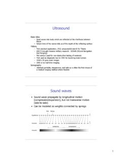
5 Higher frequency has smaller wavelength c = f Better spatial resolution Higher frequency waves degrade faster with distance Trade-off between penetration depth and spatial resolution Basics of Ultrasound Propagation of Ultrasound waves are defined by the theory of acoustics Ultrasound moves in a wavelike fashion by expansion and compression of the medium through which it travels Ultrasound waves travel at different speeds depending on material Ultrasound waves can be absorbed, refracted, focused, reflected, and scattered. Basics of Ultrasound Process Overview Transducer (electrical signal a acoustic signal) generates pulses of Ultrasound and sends them into patient Organ boundaries and complex tissues produces echoes (reflection or scattering)
6 Which are detected by the transducer Echoes displayed on a grayscale anatomical image Each point in the image corresponds to an anatomical location of an echo-generating structure Brightness corresponds to echo strength Echolocation Biosonar or Active navigation Animals emit sounds and listen for echoes Used to navigate or to hunt Bats, toothed whales and dolphins, shrews, and cave-dwelling birds use biosonar Ultrasound , audible, and infrasound frequencies Many other animals use passive biosonar Humans Listening is equivalent to passive biosonar Sound Localization Localization determined by interaural space Interaural time difference (ITD) Depends on head size Interaural sound level difference (ILD)
7 Head shadows Frequency dependent 20 Hz 80 Hz 800 Hz 1600 Hz 20 kHz Small phase delays Phase delays easy to detect Transition Zone Small level differences Unambiguous phase detection is difficult Level difference increases Both phase and level difference are used /2 ITD ILD f Sound Localization Why do animals communicate within different audible frequency ranges? Head size impacts audible frequency range of animals Also affected by ear position and movement Larger animals utilize lower frequencies Larger animals communicate over longer distances Lower frequencies have less acoustic loss with distance Smaller animals need to resolve smaller objects Higher frequencies have better spatial resolution Sound Localization Range Detection Pulse-echo ranging Localization Difference in arrival time to ears Difference in sound level between ears Interaural space R=c t2 x=c t2 Sonar SOund Navigation And Ranging Distance Pulse-echo ranging Bearing Similar to
8 Localization Relative arrival times measured multiple hydrophones or array Speed Doppler effect Difference in frequency between transmitted and received signals Converted to velocity Speed of transmitter must be accounted for Several sonar beams used Sonar Pulse-echo ranging Uses of Ultrasound Ultrasound Imaging / Detection Medical Sonography 3-20 MHz Sonar Hz kHz range Non-destructive testing (NDT) kHz low MHz range Detection of cracks in materials Medical sonography Non-destructive testing Uses of Ultrasound Monitoring Structural Health Monitoring Long term damage detection Infrastructure, aircraft Embedded sensor networks kHz-low MHz range Fetal Heart Monitoring Continuous detection and monitoring of fetal heart beat Low MHz range Sensor network for SHM Damage detection with sensor network External fetal heart monitoring Uses of Ultrasound Ablation/Destruction of Tissues Lithotripsy ablation of kidney stones Uterine fibroids (FDA approved)
9 Tumor ablation MRI or Ultrasound guided Ultrasound Hyperthermia Treatment Low level heating (<45 C) Combined with radiation/chemotherapy Other Uses Drug activation (Focused heating of drugs) Vibration (Wire bonding) Tissue cutting and hemostasis (Harmonic scalpel) Water treatment Focused Ultrasound Lithotripsy Physics of Ultrasound Compressional (Longitudinal) Waves Oscillation occurs in direction of wave propagation Zones of compression & rarefaction Speaker analogy Used in medical sonography Shear (Transverse) Waves Oscillation occurs normal to direction of wave propagation Cork bobbing in water analogy Only supported by hard tissues Important in transducer design and NDT Wave Propagation Surface (Rayleigh) Waves Travel along surfaces of hard materials, up to 1 depth Elliptical motion, combines compressional & shear motion Lamb (Guided) Waves Travel within thin plates or layers Important in NDT Wave Propagation Wave Propagation Animation Animation courtesy of Prof.
10 Daniel Russell, Penn State University , and Compressional or Longitudal Wave Shear or Transverse Wave Rayleigh or Surface Wave Lamb or Guided Wave Anatomy of a Wave Amplitude Change in magnitude Units of pressure (Pa or N/m2) Wavelength ( ) One complete wave cycle Unit of distance (m) , thicknesses are important in acoustics Constructive/Destructive interference Frequency (f) Number of vibrations that a molecule makes/second Unit of cycles/s (Hz) Period (T = 1/f) Elapsed time between compression zones Units of time (s) Wavelength Amplitude Speed of Sound Often called velocity , but is a scalar value Depends on medium Air = 330 m/s Water = 1480 m/s Average Soft Tissue = 1540 m/s Bone = 4080 m/s Steel = 5960 m/s Speed of sounds varies slightly with Temperature Water = 1480 m/s at 20 C Water = 1570 m/s at 37 C Depends on elasticity of the material through which it travels Particle velocity velocity of individual oscillating particles Phase velocity Rate at which the phase of the wave propagates in space Speed of any one frequency component Speed of Sound Wave propagation Pressure waves travel thru a medium at a frequency range of 3-20 MHz in medical ultrasonography