Transcription of Univers™ II Total Shoulder System - University of Washington
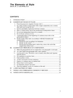
1 Total Shoulder System Anatomic adaptability .. simplified SURGICAL TECHNIQUE. Inclination Version Offset 125 - 140 +/- 10 mm A IIof Possibilities I I. The Univers II Total Shoulder System was designed in cooperation with Anthony Romeo, , Rush University Medical Center, Chicago, IL. and Professor Peter Habermeyer, , ATOS Clinic, Heidelberg, Germany. II Total Shoulder System . I I. The Definitive Anatomic Solution for Total Shoulder Arthroplasty IMPLANT DESIGN RATIONALE. The Univers II humeral component was designed to account for anatomical variations of the proximal humerus commonly encountered by the surgeon. Variable adjustment with respect to the inclination angle, version and head offset are features critical to reconstruction of the proximal humerus.
2 The simplified design of the Univers II humeral component allows the surgeon to adapt the humeral stem and articular surface to the position that best represents the patient's normal anatomy. All of the adjustments can be made intraoperatively with the implant in the humeral canal. This unique feature allows the surgeon to more accurately recreate the normal anatomical relationships of the Shoulder joint. With anatomic restoration of the humerus and glenoid, soft tissue balancing of the rotator cuff is more accurate, allowing for improved functional outcome. IMPLANT FEATURES: Variable inclination, version and offset Package-to-canal design: Anatomic restoration in situ Eccentric humeral heads Multiple head diameters and heights for precise anatomic reconstruction Instruments and trays designed to maximize efficiency in the operating room Keeled and pegged glenoid options available Keeled Glenoid: Pegged Glenoid: Dual fenestrations for enhanced anchoring Unique 2-pegged design features a Reverse barbs for expansion effect within curved keel with reverse barbs and the glenoid vault large fenestration for excellent fixation strength 1.
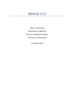
3 SURGICAL approach As described by Anthony Romeo, , Rush University Medical Center, Chicago, IL. 1. Patient Positioning Following general anesthesia, the patient is placed in the beach chair (semi-sitting) position. Typically the angle is 30-45 of elevation with respect to the operating room floor. The head and neck are secured using a ring headrest which is helpful for maintaining the head and neck position throughout the procedure. The endotracheal tube and intravenous lines are positioned on the contralateral side of the affected Shoulder . The upper body is brought to the edge of the operating room table to allow full extension of the arm, which is essential for the exposure of the proximal humerus.
4 A folded towel is placed behind the medial border of the scapula to stabilize the position of the glenoid throughout the procedure. A sterile preparation and draping is performed on the Shoulder and arm to allow full exposure and free movement of the entire limb. 2. Incision and EXPOSURE. The deltopectoral incision begins at the inferior border of the midsection of the clavicle, proceeds at an angle past the medial aspect of the coracoid prominence, and ends at the superior aspect of the axillary fold. The skin incision often lies directly over the cephalic vein, and therefore the interval between the deltoid and pectoralis major muscles.
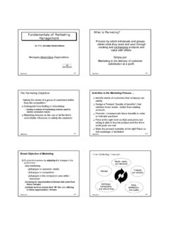
5 The cephalic vein clearly defines the junction between the deltoid and pectoralis major muscles. If the vein is not readily identified, the prominence of the coracoid also marks the deltopectoral interval, or the surgeon can identify the fibrous portion of the superior aspect of the pectoralis tendon at the distal part of the incision. The vein can be mobilized medially or laterally. The conjoined tendon complex consisting of the short head of the biceps and the coracobrachialis muscle is identified. The muscular portion of the biceps (red) is the most lateral part of the conjoined tendon, with the tendinous portion (white) just medial to the visible muscle.
6 The approach through the clavipectoral fascia is just lateral to the red stripe . representing the muscular portion of the short head of the biceps. The deltoid muscle is carefully mobilized laterally and protected. The coracoacromial ligament is maintained (not released). A thin retractor (eg. Homann or Darrach) is placed under the coracoacromial ligament to provide exposure to the superior aspect of the subscapularis and humerus. Retraction of the deltoid and pectoralis major is maintained with a self-retaining retractor. Frequently, the superior 1-2 cm of the pectoralis tendon is released to provide exposure to the inferior aspect of the subscapularis and the anterior circumflex vessels.
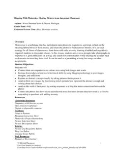
7 The arm is then externally rotated to further expose the boundaries of the subscapularis muscle and tendon insertion. The superior aspect of the subscapularis tendon is at the level of the coracoid tip and can be clearly identified by excising part of the subcoracoid bursa and rotator interval capsule. The inferior border of the subscapularis tendon is at the level of the anterior circumflex vessels. This group of vessels includes the anterior circumflex artery bordered by two anterior circumflex veins and is commonly referred to as the Three Sisters . The lateral border of the subscapularis tendon is identified just medial to the bicipital groove.
8 Two tag stitches using #2 FiberWire are placed into the medial aspect of the subscapularis tendon in preparation for the subscapularis release. 3. Subscapularis Release For uncomplicated Shoulder arthroplasty a transtendon approach is preferred, leaving 5 mm of tendon attached to the lesser tuberosity for later repair of the tendon to both the bone and the remaining tendon. The subscapularis tendon is entirely released beginning at the rotator interval over the top of the biceps and proceeding inferiorly below the level of the anterior circumflex vessels, continuing along the humeral neck. The humerus is externally rotated to facilitate the release of the capsule from the humerus to the 6 o'clock position on the humerus.
9 2. 4. Glenohumeral Capsule Release Once the subscapularis tendon is released from the humerus, the surgeon has an opportunity to release the anterior and inferior capsule with excellent direct visualization. This capsular release is a routine part of Shoulder arthroplasty for patients with a loss of external rotation, most commonly seen in osteoarthritis patients. A ring retractor (Fukuda) is placed across the glenohumeral joint and hooked on the posterior glenoid. The retractor is used to sublux the humerus, posterior and lateral, placing tension in the inferior capsule. The junction between the muscular portion of the subscapularis (red) and the capsule (white) is clearly identified.
10 The axillary nerve is generally just inferior to the muscular portion of the subscapularis or less than 1 cm from the capsule. The nerve should be identified and protected. With tension in the capsule, it is released from lateral to medial, ending at the 6 o'clock position on the glenoid. The anterior capsule is bluntly separated from the subscapularis and sharply incised (capsulotomy). Finally, the fibrous attachments from the lateral aspect of the coracoid to the subscapularis are released completing mobilization of the subscapularis when combined with the anterior/inferior capsulotomy. The release should remain lateral to the coracoid process to avoid injury to the nerve of the subscapularis and the brachial plexus.