Transcription of Updated 2017 ICO Guidelines for Diabetic Eye Care
1 ICO Guidelines for Diabetic Eye CareUpdated 2017 International Council of Ophthalmology | Guidelines for Diabetic Eye care | Page iCopyright ICO January 2017. Translation and adaption for non-commercial local use is encouraged, but please credit Guidelines for Diabetic Eye CareThe International Council of Ophthalmology (ICO) developed the ICO Guidelines for Diabetic Eye care to serve a supportive and educational role for ophthalmologists and eye care providers worldwide. They are intended to improve the quality of eye care for patients around the Guidelines address the needs and requirements for the following levels of service: High Resource Settings: Advanced or state-of-the-art screening and management of DR based on current evidence, and clinical trials.
2 Low-/Intermediate Resource Settings: Essential or core to mid-level service for screening and management of DR with consideration for availability and access to care in different Guidelines are designed to inform ophthalmologists about the requirements for the screening and detection of Diabetic retinopathy, and the appropriate assessment and management of patients with Diabetic retinopathy. The Guidelines also demonstrate the need for ophthalmologists to work with primary care providers and appropriate specialists such as diabetes and Diabetic retinopathy a rapidly increasing problem worldwide, it is vital to ensure that ophthalmologists and eye care providers are adequately ICO believes an ethical approach is indispensable, as it is the first step toward quality clinical practices.
3 Download the ICO Code of Ethics at: (PDF 198 KB). The Guidelines are designed to be a working document and will be Updated on an ongoing basis. They were first released in December 2013. This document was reviewed, revised, and Updated in 2016. The ICO hopes these Guidelines are easy to read, translate, and adapt for local use. The ICO welcomes any feedback, comments, or email us at: 2013 Task Force on Diabetic Eye care Hugh Taylor, MD, AC, Chairman Susanne Binder, MD Taraprasad Das, MD, FRCS Michel Farah, MD Frederick Ferris, MD Pascale Massin, MD, PhD, MBA Wanjiku Mathenge, MD, PhD, MBChB Serge Resnikoff, MD, PhD Bruce E.
4 Spivey, MD, MS, MEd Juan Verdaguer, MD Tien Yin Wong, MD, PhD Peiquan Zhao, MD2016 Diabetic Eye care Committee Tien Yin Wong, MBBS, PhD (Singapore), Chairman Lloyd Paul Aiello, MD, PhD (USA) Frederick Ferris, MD (USA) Neeru Gupta, MD, PhD, MBA (Canada) Ryo Kawasaki, MD, MPH, PhD (Japan) Van Lansingh, MD, PhD (Mexico) Mauricio Maia, MD, PhD (Brazil) Wanjiku Mathenge, MD, PhD, MBChB (Rwanda) Sunil Moreker, MBBS (India) Mahi Muqit, FRCO phth, PhD (UK) Serge Resnikoff, MD, PhD (Switzerland) Paisan Ruamviboonsuk, MD (Thailand) Jennifer Sun, MD, MPH (USA) Hugh Taylor, MD, AC (Australia) Juan Verdaguer, MD (Chile) Peiquan Zhao, MD (China)International Council of Ophthalmology | Guidelines for Diabetic Eye care | Page iiCopyright ICO January 2017.
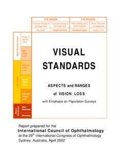
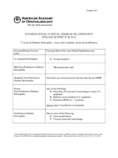
5 Translation and adaption for non-commercial local use is encouraged, but please credit Epidemiology of Diabetic Retinopathy Classification of Diabetic Retinopathy Nonproliferative Diabetic Retinopathy (NPDR) Proliferative Diabetic Retinopathy (PDR) Diabetic Macular Edema (DME) Table 1. International Classification of Diabetic Retinopathy and Diabetic Macular Edema Table 2a. Re-examination and Referral Recommendations Based on Simplified Classification of Diabetic Retinopathy and Diabetic Macular Edema for High Resource Settings Table 2b.
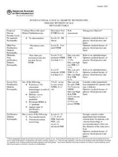
6 Re-examination and Referral Recommendations Based on Simplified Classification of Diabetic Retinopathy and Diabetic Macular Edema for Low-/Intermediate Resource SettingsScreening Guidelines Screening Guidelines Referral GuidelinesDetailed Ophthalmic Assessment of Diabetic Initial Patient Assessment Patient History (Key Elements) Initial Physical Exam (Key Elements) Fundus Examination Assessment Methods Follow-up Examination of Patients with Diabetic Retinopathy Follow-up History Follow-up Physical Exam Ancillary Tests (High Resource Settings) Patient Education Table 3a.
7 Follow-up Schedule and Management based on Diabetic Retinopathy Severity for High Resource Settings Table 3b. Follow-up Schedule and Management based on Diabetic Retinopathy Severity for Low-/Intermediate Resource Settings Treatment of Diabetic High Resource Settings Low-/Intermediate Resource Settings Panretinal Photocoagulation (PRP) Pre-treatment Discussion with Patients Lenses for PRP Table 4. Laser Spot Size Adjustment Required for Different Lenses Contact Technique for PRP Table 5. The burn characteristics for panretinal photocoagulation111112233344455666667788 88899 Table of ContentsInternational Council of Ophthalmology | Guidelines for Diabetic Eye care | Page iiiCopyright ICO January 2017.
8 Translation and adaption for non-commercial local use is encouraged, but please credit A. Technique for PRP. 13 Annex B. Recommended Practice for Intravitreal Injection. 15 Annex Table 1. Features of Diabetic Retinopathy (also see the photographs continued in the annex). 16 Annex Table 2. Features of Proliferative Diabetic Retinopathy. 17 Annex Table 3. Available Assessment Instruments and Their Advantages and Disadvantages. 17 Annex Flowchart 1. Screening for Diabetic Retinopathy. 19 Annex Flowchart 2. Treatment decision tree of DME based on central -Involvementand Vision. 19 Annex Flowchart 3.
9 Anti-VEGF treatment decision tree based on the re-treatment and follow-up schedule . 20 Figure 1. Mild nonproliferative Diabetic retinopathy with microaneurysms . 21 Figure 2. Moderate nonproliferative Diabetic retinopathy with hemorrhages, hard exudates and microaneurysms. 21 Figure 3. Moderate nonproliferative Diabetic retinopathy with moderate macular edema, with hard exudates approaching the center of the macula. 22 Figure 4. Moderate nonproliferative Diabetic retinopathy with no Diabetic macular edema. 22 Figure 5. Moderate nonproliferative Diabetic retinopathy with mild Diabetic macular edema.
10 23 Figure 6. Moderate nonproliferative Diabetic retinopathy with severe macular edema. 2356789 Treatment for Diabetic Macular Edema (DME) High Resource Settings Low-/Intermediate Resource Settings Laser Technique for Macular Edema Table 6. Modified-ETDRS and the Mild Macular Grid Laser Photocoagulation Techniques Indications for VitrectomyManagement of Diabetic Retinopathy in Special Pregnancy Management of CataractList of Suggested Indicators for Evaluation of DR ProgramsEquipmentInternational Council of Ophthalmology | Guidelines for Diabetic Eye care | Page ivCopyright ICO January 2017.