Transcription of URINARY!SYSTEM! - Mt. SAC Faculty Directory
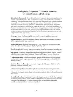
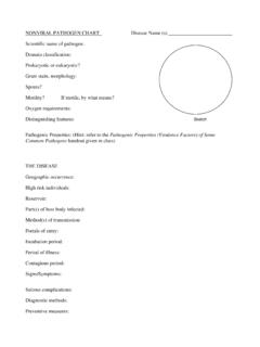
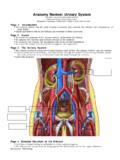
1 URINARY SYSTEM Human Anatomy Unit 3 Components Kidneys Ureters Urinary bladder Urethra Func:ons Storage of urine Bladder stores up to 1 L of urine Excre:on of urine Transport of urine out of body Regula:on: Plasma pH Blood volume/pressure Plasma ion concentra:ons (Ca2+, Na+, K+, CL- ) Assist liver in detoxifica:on, amino acid metabolism kidney Gross Anatomy Retroperitoneal Anterior surface covered with peritoneum Posterior surface directly against posterior abdominal wall Superior surface at about T12 Inferior surface at about L3 Ureters enter urinary bladder posteriorly LeT kidney 2cm superior to right Size of liver Structure of the kidney Hilum = the depression along the medial border through which several structures pass renal artery renal vein ureter renal nerves Surrounding Tissue Fibrous capsule Innermost layer of dense irregular CT Maintains shape, protec:on Adipose capsule Adipose ct of varying thickness Cushioning and insula.
2 On Renal fascia Dense irregular CT Anchors kidney to peritoneum & abdominal wall Paranephric fat Outermost, adipose CT between renal fascia and peritoneum Frontal Sec:on of the kidney Cortex Layer of renal :ssue in contact with capsule Renal columns parts of cortex that extend into the medulla between pyramids Medulla Striped due to renal tubules Renal pyramids 8- 15 present in medulla of adult Conical shape Wide base at cor:comedullary junc:on Flow of Filtrate/Urine Collec:ng ducts Collect from mul:ple nephrons Minor calyx Collect from each pyramid Major calyx Collect from minor calyx Renal pelvis Collects from calyces, passes onto Ureter Collects from pelvis Urinary Bladder Collects from ureters Histology Renal Cortex Renal Medulla Renal Tubules Nephron func:onal unit of the kidney .
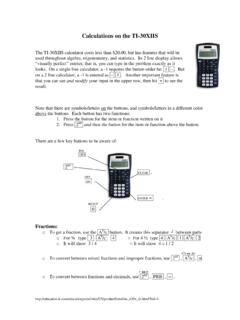
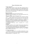
3 Each kidney contains approximately 1 million nephrons Form urine by filtering and adjus:ng composi:on of blood carried by renal vasculature Renal Portal System Renal Portal System Histological Structure of a Nephron Renal corpuscle Glomerulus Bowman s capsule Renal tubules Proximal convoluted tubule Descending limb of LOH Loop of Henle Ascending limb of LOH Distal convoluted tubule Collec:ng duct Associated blood vessels - Peritubular capillaries - Vasa recta Types of Nephrons The Glomerulus Bowman s capsule Glomerulus Afferent arteriole Efferent arteriole Podocytes The Collec:ng System Collec:ng ducts receive filtrate from DCT of nephrons Collec:ng ducts empty into minor calyces Minor calyces empty into major calyces Major calyces drain into renal pelvis Renal pelvis drains into ureter Now urine The Ureters Expandable tubes that exit the renal pelvis 3 walls Mucosa Transi:onal epithelium Muscularis smooth muscle layer Adven::a protec:ve fibrous CT Ureters drain into the posterior por:on of the urinary bladder The Urinary Bladder Func:ons to store urine Structure Rugae macroscopic folds as in the stomach flaeen when the urinary bladder is distended Trigone triangular region of the bladder no rugae loca.
4 On of openings to the ureters and urethra Histology of the Urinary Bladder Mucosal lining transi:onal epithelium Submucosa fibrous CT Muscularis detrusor muscle 3 layers of smooth muscle Serosa loose CT visceral peritoneum The Female Urethra Drains urine from urinary bladder to exterior 1- 2 inches higher risk for bladder infec:ons The Male Urethra 3 regions: prosta:c membranous penile Histology of the Urethra Mucosa varies from bladder to exterior especially in males Muscularis layer Adven::a Sphincters internal = smooth muscle (involuntary) external = skeletal muscle (voluntary)