Transcription of Version 12 May 2013 - BSAC
1 BSAC Methods for Antimicrobial Susceptibility Testing Version 12 May 2013 All enquiries to: Mandy Wootton Email: Telephone: +44 (0) 2920 746581 Version 12 January 2013 2 Contents Page Working Party members 5 Abstract 6 Preface 8 Disc Diffusion Method for Antimicrobial Susceptibility Testing 1. Preparation of plates 10 2. Selection of control organisms 11 Table 2 a Control strains to monitor test performance of antimicrobial susceptibility testing 12 2b Control strains used to confirm that the method will detect resistance 12 3. Preparation of inoculum 12 Comparison with McFarland standard 13 Preparation of the McFarland standard 13 Inoculum preparation by the growth method 13 Inoculum preparation by the direct colony suspension method 13 Adjustment of the organism suspension to the density of the McFarland standard 13 Dilution of suspension equivalent to McFarland standard in distilled water before inoculation 13 Photometric standardisation of turbidity of suspension 14 Direct susceptibility testing of urines and blood cultures 15 4.
2 Inoculation of agar plates 16 5. Antimicrobial discs 16 Storage and handling of discs 16 Application of discs 16 6. Incubation 16 Conditions of incubation 16 7. Measuring zones and interpretation of susceptibility 18 Acceptable inoculum density 18 Measuring zones 18 Use of templates for interpreting susceptibility 18 8. Oxacillin/cefoxitin testing of staphylococci 19 Detection of oxacillin resistance in Staphylococcus aureus and coagulase negative staphylococci 19 Detection of methicillin/oxacillin/cefoxitin resistance in staphylococci by use of cefoxitin as test agent 20 Interpretative tables Table MIC and zone breakpoints for: 6 Enterobacteriaceae 22 7 Acinetobacter species 27 8 Pseudomonas 28 9 Stenotrophomonas maltophilia 30 Version 12 January 2013 3 Interpretative tables continued Page 10 Staphylococci 31 11 Streptococcus pneumoniae 38 12 Enterococci 41 13 -haemolytic streptococci 43 14 -haemolytic streptococci 44 15 Moraxella catarrhalis 47 16 Neisseria gonorrhoeae 49 17 Neisseria meningitidis 51 18 Haemophilus influenzae 52 19 Pasteurella multocida 55 20 Campylobacter spp.
3 56 21 Coryneform organisms 57 22 Gram-negative anaerobes 58 23 Gram-positive anaerobes except Clostridium difficile 60 24 Clostridium difficile 63 Appendices 1 Advice on testing the susceptibility to co-trimoxazole 64 2 Efficacy of cefaclor in the treatment of respiratory infections caused by Haemophilus influenzae 65 Acknowledgment 66 References 66 Additional information 1 Susceptibility testing of Helicobacter pylori 67 2 Susceptibility testing of Brucella species 67 3 Susceptibility testing of Legionella species 67 4 Susceptibility testing of Listeria species 68 5 Susceptibility testing of topical antibiotics 68 6 Development of MIC and zone diameter breakpoints 69 Control of disc diffusion antimicrobial susceptibility testing 1 Control strains 70 2 Maintenance of control strains 70 3 Calculation of control ranges for disc diffusion 70 4 Frequency of routine testing with control strains 70 5 Use of control data to monitor the performance of disc diffusion tests 70 6 Recognition of atypical results for clinical isolates 71 7 Investigation of possible sources of error 71 8 Reporting susceptibility results when controls indicate problems 72 Table Acceptable ranges for control strains for.
4 2 Iso-Sensitest agar incubated at 35-370C in air for 18-20h 73 3 Iso-Sensitest agar supplemented with 5% defibrinated horse blood, with or without the addition of NAD, incubated at 35-370C in air for 18-20h 76 4 Detection of methicillin/oxacillin/cefoxitin resistance in staphylococci 76 5 Iso-Sensitest agar supplemented with 5% defibrinated horse blood, with or without the addition of NAD, incubated at 35-370C in 10% CO2/10% H2 /80% N2 for 18-20 h 77 6 Iso-Sensitest agar supplemented with 5% defibrinated horse blood, with or without the addition of NAD, incubated at 35-370C in 4-6% CO2 for 18-20 h 78 Version 12 January 2013 4 Page 9. Control of MIC determinations Table Target MICs for: 7 Haemophilus influenzae, Enterococcus faecalis, Streptococcus pneumoniae, Bacteroides fragilis and Neisseria gonorrhoeae 80 8 Escherichia coli, Pseudomonas aeruginosa and Staphylococcus aureus 82 9 Pasteurella multocida 84 10 Bacteroides fragilis, Bacteroides thetaiotaomicron and Clostridium perfringens 84 11 Group A streptococci 84 References 85 Suppliers 86 Useful web sites 87 Version 12 January 2013 5 Working Party Members: Dr Robin Howe (Chairman) Consultant Microbiologist Public Health Wales University Hospital of Wales Heath Park Cardiff CF14 4XW Dr.
5 Mandy Wootton (Acting Secretary) Lead Scientist Public Health Wales University Hospital of Wales Heath Park Cardiff CF14 4XW Professor Alasdair MacGowan Consultant Medical Microbiologist Southmead Hospital Westbury-on-Trym Bristol BS10 5NB Professor David Livermore Professor of Medical Microbiology Faculty of Medicine & Health Sciences Norwich Medical School University of East Anglia Norwich Research Park Norwich NR4 7TJ Dr Nicholas Brown Consultant Microbiologist Clinical Microbiology HPA Level 6 Addenbrooke's Hospital Hills Road Cambridge CB2 2QW Dr Trevor Winstanley Clinical Scientist Department of Microbiology Royal Hallamshire Hospital Glossop Road Sheffield S10 2JF Dr Derek Brown (Scientific Secretary for EUCAST) Mr Christopher Teale Veterinary Lab Agency Kendal Road Harlescott Shrewsbury Shropshire SY1 4HD Professor Gunnar Kahlmeter Central Lasarettet Klinisk Mikrobiologiska Laboratoriet 351 85 Vaxjo Sweden Dr.
6 Karen Bowker Clinical Scientist Southmead Hospital Westbury-on-Trym Bristol BS10 5NB Dr. Gerry Glynn Medical Microbiologist Microbiology Department Altnagelvin Hospital Glenshane Road Londonderry N. Ireland BT47 6SB Dr. Fiona MacKenzie Medical Microbiology Aberdeen Royal Infirmary Foresthill Aberdeen AB25 2ZN Ms Phillipa J Burns Senior BMS Microbiology Department of Medical Microbiology Manchester Medical Microbiology Partnership, HPA & Central Manchester Foundation Trust Manchester M13 9WZ All enquiries to Mandy Wootton Email: Telephone: +44 (0) 2920 746581 Version 12 January 2013 6 Abstract Summary of changes in Version 12 Table 6. MIC and zone diameter breakpoints for Enterobacteriaceae (including Salmonella, Shigella spp. and Yersinia enterocolitica) MIC and zone diameter breakpoints for Y. enterocolitica Tetracycline Table 8. MIC and zone diameter breakpoints for Pseudomonas spp.
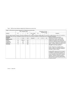
7 Note to the table Table 10. MIC and zone diameter breakpoints for staphylococci MIC and zone diameter breakpoints Levofloxacin Comments Susceptibility testing of S. saprophyticus under review Medium that should be used to test daptomycin Re-instatement of the comment for mupirocin Table 11. MIC and zone diameter breakpoints for Streptococcus pneumoniae MIC and zone diameter breakpoints Clindamycin Table 12. MIC and zone diameter breakpoints for enterococci MIC and zone diameter breakpoints Amoxicillin Table 16. MIC and zone diameter breakpoints for Neisseria gonorrhoeae Removal of disc testing recommendations Cefixime Ceftriaxone Cefotaxime Comments Use of cefuroxime screen Table 19. MIC and zone diameter breakpoints for Pasteurella multocida Changes in MIC and zone diameter breakpoints Penicillin Cefotaxime Ciprofloxacin Tetracycline Comments Disc breakpoints under review Version 12 January 2013 7 Table 20.
8 MIC and zone diameter breakpoints for Campylobacter spp. Change in MIC breakpoint Ciprofloxacin Version 12 January 2013 8 Preface Since the Journal of Antimicrobial Chemotherapy Supplement containing the BSAC standardized disc susceptibility testing method was published in 2001, there have been various changes to the recommendations and these have been posted on the BSAC website ( ). One major organizational change has been the harmonisation of MIC breakpoints in Europe. In 2002 the BSAC agreed to participate with several other European national susceptibility testing committees, namely CA-SFM (Comit de l Antibiogramme de la Soci t Fran aise de Microbiologie, France), the CRG (Commissie Richtlijnen Gevoeligheidsbepalingen (The Netherlands), DIN (Deutsches Institut f r Normung, Germany), NWGA (Norwegian Working Group on Antimicrobials, Norway) and the SRGA (Swedish Reference Group of Antibiotics, Sweden), in a project to harmonize antimicrobial breakpoints , including previously established values that varied among countries.)
9 This work is being undertaken by the European Committee on Antimicrobial Susceptibility Testing (EUCAST) with the support and collaboration of the national committees, and is funded by the European Union, the European Society for Clinical Microbiology and Infectious Diseases (ESCMID) and the national committees, including the BSAC. The review process includes application of more recent techniques, such as pharmacodynamic analysis, and current data, where available, on susceptibility distributions, resistance mechanisms and clinical outcomes as related to in vitro tests. There is extensive discussion between EUCAST and the national committees, including the BSAC Working Party on antimicrobial susceptibility testing, and wide consultation on proposals. In the interest of international standardization of susceptibility testing, and the need to update older breakpoints , these developments are welcomed by the BSAC.
10 The implication of such harmonization is that over time some MIC breakpoints will change slightly and these changes will be reflected, where necessary, in corresponding changes to zone diameter breakpoints in the BSAC disc diffusion method. It is appreciated that changes in the method require additional work for laboratories in changing templates and laboratory information systems, and that the wider use of `intermediate categories will add complexity. Nevertheless the benefits of international standardization are considerable, and review of some older breakpoints is undoubtedly warranted. In line with the European consensus EUCAST MIC breakpoints are defined as follows: Clinically resistant: level of antimicrobial susceptibility which results in a high likelihood of therapeutic failure Clinically susceptible: level of antimicrobial susceptibility associated with a high likelihood of therapeutic success Clinically intermediate: a level of antimicrobial susceptibility associated with uncertain therapeutic effect.