Transcription of Vestibular Tests & Measures: Study Guide
1 Vestibular Tests & Measures: Study Guide Nystagmus is described by the direction of the quick phase. Rotary / Torsional N. is described by the direction that the superior pole of the iris moves, L or R. Directions to perform TEST Positive sign demonstrated by Central vs. Peripheral Eye Movement Range Take your finger out past 18-24 to examine if the patient has full ocular range of motion. Ask the patient to follow moving target that is held several feet in front of the patient s face (to avoid convergence of eyes.) Combination: ocular muscles Smooth Pursuit Maintains gaze stabilization when rate of eye movement is < 60d/sec, slower then VOR Gain testing Hold the patient s head stationary. Have the patient follow your slowly moving finger horizontally (from center to 30 right and then to 30 left), and then vertically (center to 30 up to 30 down).
2 Can use an H pattern. The test can be repeated; you may have to hold the eyelids up in order to see the downward eye movement clearly. Eyes do not move synchronously or symmetrically. Central End Point Nystagmus (physiological response) During maintenance of an extreme eye position. Head fixed. Eyes follow my finger and then held at the end point. 1-2 beats is normal. Central Gaze evoked Nystagmus (abnormal response) Observe direction of nystagmus in each test position. Hold the patient s head stationary. Have the patient follow your finger so she/he is looking 30 to the right, left, up, down. Pause for 20 seconds in each of those positions to observe for nystagmus. Note the direction of the nystagmus in each position. Be sure to keep your finger 18-24 inches away from the patient s face throughout the entire test.
3 Persistent nystagmus Central Saccades Extra involuntary eye movements during horizontal and vertical tracking. Normal saccadic movement: hold the patient s head stationary. Hold your finger about 15 degrees to one side of your nose. Ask the patient to look at your nose, then at your finger, repeating several times. Do this from the right, left, up, and down. You are looking for the number of eye movements it takes for the patient s eyes to reach the target. Normal is < 2. Central Near Point Convergence (c/s glasses) Normal: diplopia at < 5cm from nose; observe for symmetrical eye movement Hold target at nose level, arm s length away. Focus on the target as you move it toward your nose. Stop when target turns double. Measure distance from nose to target.
4 Repeat 2-3 times. > 6cm = Convergence insufficiency Central Diplopia Patient report of double vision. Test both near and far field of vision. Central Oscillopsia: visual blurring with head movement Decreased VOR gaze stability with head motion Patient report of environment moving or oscillating, as they walk through it. May also be reported as seaweed movement without head mvmt. Peripheral or Central Alternate Cover Test Therapist alternately covers and uncovers each eye, while patient keeps their eyes open, focused on therapist s nose. Covered eye has moved from midline; phoria Utricle hypofunction, or brainstem pathways Cover Test Patient looks at therapist s nose. Cover one eye at a time for 2 sec. Observe the uncovered eye for movement.
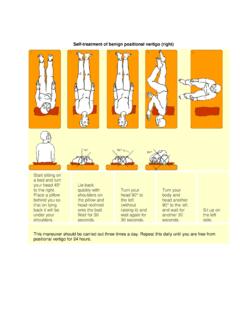
5 Ocular misalignment; eye readjusts when opposite eye covered; tropia / strabismus Ocular muscle dysfunction Ocular Tilt Reaction (OTR) (can accompany Wallenberg syndrome) Observation: Triad = head tilt + skew deviation + torsion See illustration of OTR in: O Sullivan 6th ed. , Fig. (O Sullivan 5th ed. , Fig. ) Unilateral brainstem , Medullary infarct Subjective Visual Vertical (SVV); Subjective Visual Horizontal (SVH) Equipment: 5 gallon bucket with a straight line drawn across the bottom of the bucket (inside and outside); inclinometer on outside Task: with their head inside the bucket, therapist turns the bucket until patient perceives the line to be vertical; repeat twice.
6 Test horizontal direction Abnormal if > 2 degrees off Peripheral or Central: Otoliths to Vestibular cortex Spontaneous Nystagmus (not movement or position related) May indicate an acute Vestibular dysfunction Holding the patient s head with one hand. Have the patient look straight ahead without focusing, observe for nystagmus. Horizontal Nystagmus that stops w gaze fixation = Peripheral Nystagmus that does NOT stop with gaze fixation = Central Optokinetic Nystagmus (normal physiological occurrence of nystagmus under these conditions) Nystagmus should beat in opposite direction to the target s movement Using a vertical bar chart, tell the patient to stare at the line that is in front of you , while you slowly move the chart in one direction. Repeat this procedure to the opposite direction.
7 Observe for nystagmus. No response, or atypical eye movement. Central VOR Gain 1. Maintained Fixation Maintains fixed gaze with head movement (eyes move opposite to head). Keep your eyes on my finger. Move your head quickly side to side; repeat up & down Done at a rate of > 60 / sec. (faster than smooth pursuit) VOR x 1 dizziness , nausea, headache. Not able to maintain focus on target. Vestibular hypofunction 2. Head Impulse Test. aka Head Thrust Test (eyes open: EO) The patient will need to understand what will be done so their neck is relaxed during the test. If you noted that the patient had pain or significant restriction in cervical spine mobility, this test should be performed with extreme caution or should be deferred. Grasp the patient s head with both hands on the side of their head.
8 Flex neck down 30 so that horizontal semi-circular canals are level in the horizontal plane. Instruct the patient to look at your nose. Move the patient s head slowly back and forth being sure the patient is Saccade (to catch up) is a positive sign. A positive head thrust to the L side indicates a L lesion Peripheral dysfunction with corrective saccades. relaxed. Then, suddenly move the patient s head in one direction and stop. The horizontal head movement should be moved through a small amplitude with the position held at the end. Observe for the patient s ability to maintain visual fixation. You should note if the patient makes corrective saccades to re-fixate on your nose and the direction of head movement that caused the re-fixation saccades, if a thrust to the L yields a saccade to re-fixate on your nose a Left peripheral lesion is indicated.
9 Note: If you are uncomfortable moving the person s head from center to an eccentric position, try moving the person s head from an eccentric position to center This test helps differentiate between L or R or bilateral lesion. 3. Head Shake Test. (eyes closed: EC) Velocity storage integration; dynamic balance of labyrinths Eyes are closed and with 30 neck flexion (horizontal SCC position). Therapist shakes their head side to side at 2 Hz (200-240 bpm) for 20 cycles. Stop and then they open their eyes. Observe for nystagmus. Nystagmus; fast phase to intact side Peripheral UVL 4. VOR Cancellation (VOR Suppression) Cerebellum inhibits VOR Gain during VOR Cancellation Maintain fixation with head movement Therapist holds target in front of subject at eye level.
10 Ask subject to move head and eyes to follow the target as the therapist moves the target slowly side to side, up and down, and in diagonals. Cue: Pretend there is a string from your nose to my finger/target . The arc of movement should be within 30 of the midline in all directions. Saccades, Nystagmus, Difficulty crossing midline. Central: Cerebellar 5. Dynamic Visual Acuity Test (DVA) Test of functional VOR. STATIC TEST: Have the patient wear their glasses if they need distance correction. Depending on the type of acuity chart being used, have the patient sit the appropriate distance from the chart. (The ETDRS charts are designed to be viewed from a distance of 10 feet to provide Snellen equivalent acuity ratios or LogMAR values as noted on the chart).