Transcription of Veterinary Developmental Anatomy
1 12013 VeterinaryDevelopmental Anatomy ( Veterinary Embryology) CVM 6903byThomas F. Fletcher, DVM, PhDand Alvin F. Weber, DVM, PhD2 CONTENTSE arly Embryogenesis ..3 Musculo-Skeletal Development ..16 Serous Body Cavities ..23 Cardiovascular System ..25 Digestive System ..32 Respiratory System ..38 Urinary System ..41 Genital System ..44 Pharynx, Face, Nasal Cavity & System & Special Senses ..56 Appendix I. Gametogenesis ..69 Appendix II. Mitosis and Meiosis ..71 Appendix III. List of Anomalies ..753 Early EmbryogenesisEmbryogenesis: formation of body structures & organs (organogenesis) requires cell division (proliferation) and cell differentiation (specialization) produces the great variety of cell types and extracellular products found in the specialization: selective gene expression (and resultant protein production) is the ultimate explanation for the cell differentiation process during embryogenesis.
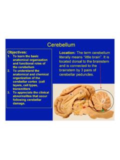
2 Genetic expression by a particular cell depends on the cell s previous genetic history (com-mitment lineage) and its current cellular environment (intercellular communications).Cell differentiation is the result of cells expressing some genes and suppressing others within a common genome. Cells differ because they produced different & peptides are: structural components (cytoskeleton or extracellular structures) enzymes (controlling cell metabolism) secretory products ( , hormones; digestive enzymes; etc.) channels & pumps (passage of molecules across membranes) receptors (communication, etc.)Periods: Embryonic Period defined as the time from fertilization to the earliest (primordial) stages of organ development (about 30 days in dog, cat, sheep, pig; almost 60 days in horse, cattle, human).
3 Fetal Period the time between the embryonic period and parturition (the end of gesta-tion), during which organs grow and begin to function. Fertilization: union of a haploid oocyte and a haploid spermatozoon, producing a diploid zygote (a pleuripotent cell capable of developing into a new individual) fertilization begins with gamete fusion (zygote formation) fertilization ends with the initiation of zygote cell division (the start of cleavage) Fertilization related details: fusion of a spermatozoon with an oocyte takes place in the uterine tube, near the ovary the spermatozoon must bind to a specific glycoprotein on the zona pellucida surrounding the oocyte [this species recognition process prevents union with foreign sperm];specialized cellstellate neuron, cell committed cells neural epitheliumneuroblastglioblastpyramidal , Cell Differentiation4 then the spermatozoon releases degradative enzymes (acrosomal reaction) [the enzymes denature the zona pellucida, allowing the sperm cell to penetrate the barrier] spermatozoon and oocyte plasma membranes fuse (secondary oocyte completes meiosis) the oocyte immediately cancels its membrane potential (via Ca++ influx) and then denatures its zona pellucida (via enzymes are released by exocytosis from oocyte cytoplasmic granules) [this prevents fusion by additional sperm] male & female haploid pronuclei make contact, lose their nuclear membranes, and begin mitosis (mitosis begins 12 hours after sperm fusion.)
4 DNA synthesis takes place before mitosis) Oocyte (enveloped by a zona pellucida (glycoprotein membrane) and corona radiata (granulosa cells) at ovulation) selective follicles mature at each cycle (in response to circulating FSH hormone from the pituitary) oogonia (germ cells) give rise to primary oocytes by mitosis within the embryo primary oocytes initiate Meiosis I (reduction division ) within the embryo and only resume Meiosis I following ovulation (being suspended in Meiosis I by inhibitory secretion of follicle granulosa cells) secondary oocytes complete meiosis (Meiosis II) following fertilization (if unfertilized they degenerate), producing a fertilized oocyte (ovum). Spermatozoa (several hundred million per ejaculate) propelled from vagina to uterine tube by contraction of female genital tract spermatogonia (germ cells) give rise to primary spermatocytes by mitosis repetitively following puberty primary spermatocytes undergo Meiosis I (reduction division ) producing secondary spermatocytes secondary spermatocytes complete meiosis (Meiosis II), producing spermatids that undergo transformation into spermatozoa (spermiogenesis) subsequently, spermatozoa undergo capacitation (removal of surface proteins that would impede contact with an oocyte)Cleavage: refers to the initial series of mitotic divisions by which the large zygote is fractionated into numerous normal size cells.
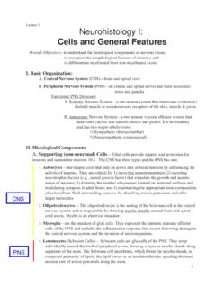
5 Each daughter cell of the cleavage process is termed a blastomere. cleavage begins with a zygote, progresses through compaction to a morula stage and terminates at the start of the blastocyst (blastula) stage the first eight blastomeres are undifferentiated and have identical potential in mammals; thereafter, blastomeres differentiate into inner & outer cells with different missionsNote: The first cleavage division occurs 1 to 5 days following ovulation (depending on species), thereafter cells divide about once every 12 hours; As many as eight generations of mitoses may occur without intervening cell growth (cytoplasmic increase). Thus, , one 150 micron diameter zygote can becomes a collection of 256 cells, each about 7 microns in DivisionSecondCleavage DivisionMorulaBlastula(Blastocyst)zonape llucidablastomeresouterblastomeresinnerb lastomerestrophoblastsblastocoeleinnerce ll mass5 Morula [ small mulberry] a solid ball of blastomeres within a zona pellucida (typically consisting of 16 to 64 blastomeres) blastomeres become compacted; cells on the inside differentiate from those along the surface of the morula: outer blastomeres become flattened and form tight junctions (reducing fluid permeability); they develop the capacity to secrete fluid (internally).
6 They are destined to become trophoblasts which form the chorion & amnion (fetal membranes) of the conceptus; inner blastomeres form gap junctions to maximize intercellular communication; they are destined to become inner cell mass which forms the embryo itself (plus two fetal membranes).Note: As few as three inner blastomeres are sufficient to produce an entire embryo (and adult). When a morula leaves the uterine tube and enters the uterus (uterine horn) it is at about the 16-cell stage, around 4 to 7 days after fertilization (depending on species). The 32-cell stage morula (5-7 days post ovulation) is ideal for embryo transfer in (or Blastula) develops during the second week, after the zona pellucida ruptures consists of a large number of blastomeres arranged to form a hollow, fluid-filled, spherical or cylindrical structure contains an inner cell mass (embryoblast), evident as a collection of cells localized inside one polar end of the blastula surface cells of the blastocyst are designated trophoblasts (future chorion of the conceptus) the cavity of the blastocyst is called a blastocoele eventually the blastocyst attaches to or implants within the uterine wall (pending species).
7 Cleavage in fish, reptiles, and birds: Large quantities of yolk impede cell division during cleavage. Thus a blastodisc (rather than a spherical or elliptical blastocyst) is formed at the animal pole of the egg. A telolecithal ovum (egg with large amounts of asymmetrically distributed yolk) has an animal pole where the nucleus is located and an opposite vegetal pole where yolk is concentrated. Cleavage is par-tial (meroblastic): cells divide more rapidly at the animal pole than at the vegetal pole, resulting in many, small blastomeres at the animal pole and a few, large macromeres at the vegetal pole. In contrast, mammalian ovum has meager amounts of yolk (oligolecithal ovum) which is uni-formly distributed (isolecithal). Cleavage is holoblastic (total) and each blastomere division produces two equal-size daughter cells.
8 Thus animal and vegetal poles are not evident in mammalian Monozygotic: identical (same genetic composition) twins can result from either: 1] separation of early blastomeres (up to the 8-cell stage) each of the separate blastomere(s) develops into an independent conceptus; or 2] separation of inner blastomeres within a single morula each of the separate blastomere(s) develops into an independent embryo and both embryos share a common placenta (this is less common than the first possibility). Note: Separations later in embryonic development result in conjoined twins (diplopagus; Siamese twins), or double heads, etc. types of : fraternal twins result when two zygotes develop independently during the same pregnancy (independence can be compromised by fusion of fetal membranes and blood supplies).
9 It is possible for fraternal blastomeres to merge and produce a single conceptus with two different genotypes (a chimera).6 GERM LAYERSE ctoderm, mesoderm and endoderm are designated primary germ layers because origins of all organs can be traced back to these three forms epidermis of the skin, epithelium of the oral and nasal cavities, and the nervous system and sense organs. Mesoderm forms muscle and connective tissue, including bone, and components of the circulatory, urinary and genital systems. Endoderm forms mucosal epithelium and glands of respiratory and digestive : The morphogenic process that gives rise to three germ layers: ectoderm, mesoderm, and endoderm. (In some species, evidence of primitive gut formation can be seen [gastrula little stomach].)
10 Gastrulation includes the following sequence, beginning with a blastocyst: A thickened embryonic disc becomes evident at the blastocyst surface, due to cell prolifera-tion of the inner cell mass cells. Trophoblast cells overlaying the inner cell mass degenerate in domestic mammals (in the mouse and human, trophoblast cells overlaying the inner cell mass separate and, instead of degenerating, become amnionic wall.) From the inner cell mass, cells proliferate, break loose (delaminate), and migrate to form a new cell layer inside the trophoblast layer. The new layer of cells, called the hypoblast, will form a yolk sac. The remaining inner cell mass may be called the epiblast. On the epiblast surface, a primitive streak forms as differential cell growth generates a pair of ridges separated by a depression.