Transcription of What are neurofibromas? Who gets neurofibromas?
1 What are neurofibromas ? neurofibromas are benign (not malignant) tumors/growths of nerves. They are basically overgrowth and disorganization of normal nerve tissue with the addition of inflammatory cells and blood vessels. neurofibromas are made of nerve fibers, Schwann cells (cells that cover the nerve fibers), blood vessels, inflammatory white blood cells (mast cells), and connective tissue (fibroblasts and loose material called extra cellular matrix). The mast cells can release histamine which causes the neurofibromas to itch or hurt. Who gets neurofibromas ? neurofibromas are a common tumor. Not everyone who has a neurofibroma has NF. People with NF1 usually get multiple neurofibromas .
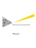
2 In neurofibromas , the nerve fibers run through the tumor. This means that removing the neurofibroma always means cutting the nerve (see treatment below). The diagram to the right shows a nerve that is swollen or ballooned out by a neurofibroma (in pink). It is compared to a Schwannoma which is a tumor of the covering of the nerve so that the tumor (in pink) simply lies on top of the nerve. Types of neurofibromas in NF1 In NF1 we see: Diffuse neurofibromas Cutaneous or Dermal neurofibromas Intramuscular neurofibromas Plexiform neurofibromas Diffuse neurofibromas are tumors that involve the full thickness of the skin (from the surface all the way down to the base of the skin (fascia).)
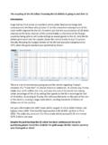
3 Diffuse neurofibromas stop at the fascia and do not usually go any deeper. Diffuse neurofibromas are uncommon. They are often seen in the scalp and feel soft, almost squishy and they do not have clear margins (you can t easily tell where the tumor stops). You can also see diffuse neurofibromas on the trunk (often protruding out like a love handle ) and they are usually associated with diffuse hyperpigmentation so that they look like a very large squishy caf au lait spot. Diffuse neurofibromas are present in early childhood and can grow. Picture of a diffuse neurofibroma of the shoulder Cutaneous or Dermal neurofibromas are the most common type of neurofibroma. These are the bumps or lumps that occur on the skin.
4 They usually start in teenage years or young adults and rarely start in childhood. They increase in size and number over the years. Sometimes they don t start until middle age. They have clearly defined borders and can be removed if necessary. The number of skin tumors that someone has varies tremendously. Some patients have very few cutaneous neurofibromas . Some patients have very large numbers of cutaneous neurofibromas . It is less common to have a very large number and the tendency for very large numbers of them may run in families. Intramuscular neurofibromas are tumors that occur in the muscle. They are usually growths along very small nerves. Sometimes they cause pain and usually they can be removed (although removing them leaves a scar).
5 Sometimes intramuscular neurofibromas are actually plexiform neurofibromas that occur in a chain or network. Plexiform neurofibromas are made up of the same cells as dermal or cutaneous neurofibromas and occur in about half of all people with NF1. Plexiform neurofibromas tend to be tumors of large nerves and are often internal, but they can also involve small nerves and the superficial skin. Plexiform neurofibromas differ from cutaneous neurofibromas in that they have more connective tissue (extracellular matrix) that separates the nerve fibers. Plexiform neurofibromas tend to be congenital meaning we think patients are born with them, but they may not cause any problems and they may not be apparent for years, if ever.
6 Plexiform neurofibromas can grow. They are more likely to grow in childhood but they can also grow in adult years. There is no evidence that they get worse with puberty. Plexiform neurofibromas have different subtypes. Types of Plexiform Tumors When plexiform neurofibromas involve the skin they usually extend through the skin and extend below the skin and fascia into the muscle. This type of plexiform neurofibroma may be referred to as a diffuse plexiform neurofibroma because it does not have clear margins and tends to have little fingers of tumor that invade the muscle or other tissue. When patients with diffuse plexiform neurofibromas are infants the tumors may not be easy to see or sometimes , if there is hyperpigmentation with it, it may just look like very large caf au lait spots.
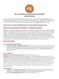
7 However, slowly the skin may thicken and enlarge and distort the normal appearance of the part of the body that is affected. To the right is a picture of a child with a diffuse plexiform neurofibroma of the face. You can see that one side of the face looks different than the other and is a little swollen. That is because the skin and some of the deep tissue of one side of the face has a diffuse plexiform neurofibroma. However, you can t really see any tumor or a clear margin or point where the changes begin or stop. Picture of patient with a few cutaneous neurofibromas of the arm Picture of a child with a diffuse plexiform neurofibroma of the face. Below is an MRI through the legs of a patient with a diffuse plexiform neurofibroma of one leg.
8 You can see how one leg is bigger (the right side of the picture) compared to the other leg and how the muscle fibers (the light colored areas) are not as dense because they are teased apart by the plexiform neurofibroma, but there is no obvious tumor no clear margins for what is normal and what is abnormal. Plexiform neurofibromas often involve the nerves coming off the spinal cord. They can also involve large nerves like the sciatic nerve of the leg. They tend to enlarge or thicken the nerve that is affected and sometimes appear to produce little clusters of tumors along the nerve. We call this type of plexiform neurofibroma a nodular plexiform neurofibroma. The picture on the right shows the legs of a patient.
9 There are a number of small white spots in the leg that are intramuscular plexiform neurofibromas and two long white cord like structures that are the sciatic nerves. The sciatic nerves are thick and have a cluster of tumors along them. What kinds of problems occur with neurofibromas ? Most neurofibromas grow and growth may or may not cause problems. neurofibromas are often itchy (because the mast cells present in the neurofibromas release histamine) and they can hurt (as a sign of growth or when they are touched or injured). neurofibromas can grow after trauma or a simple injury to the neurofibroma. The trauma or injury can damage the blood vessels associated with the neurofibroma and sometimes this results in very sudden swelling of the tumor.
10 If neurofibromas hurt you should discuss that with your doctor. Severe pain can also be a warning sign that a part of the tumor is becoming malignant (cancerous). Malignant deterioration of a plexiform neurofibroma happens in about 9% of patients with NF1. It is a very serious complication that requires immediate attention. If malignant deterioration of a plexiform neurofibroma occurs, it is usually in late teenage or adult years. Most neurofibromas , including plexiform neurofibromas don t hurt. Diffuse neurofibromas can also become malignant. Cutaneous neurofibormas never become malignant. You should also alert your doctor to growth of your cutaneous neurofibromas . Sudden growth of tumors could be a sign of bleeding in the tumor and sometimes if the tumor is a plexiform neurofibroma or diffuse neurofibroma, the bleeding can be severe and needs to be dealt with emergently.