
Electrocardiogram (EKG) Interpretation
1. Isovolumetric ventricular contraction: With ventricular depolarization, pressure increases in the ventricles and the tricuspid and mitral valves close while the pulmonic and aortic valves remain closed as well. 2. Ventricular ejection: The pulmonic and aortic valves open, and the ventricles eject blood (ventricular systole). 3.
Download Electrocardiogram (EKG) Interpretation

Information
Domain:
Source:
Link to this page:
Documents from same domain
Medical Record Documentation and Legal Aspects …
www.rn.orgMedical Record Documentation and Legal Aspects WWW.RN.ORG® Reviewed May, 2017, Expires May, 2019 Provider Information and Specifics available on our Website

Cultural Competence in Healthcare - - RN.org®
www.rn.orgCultural Competence in Healthcare WWW.RN.ORG® Reviewed May, 2017, Expires May, 2019 Provider Information and Specifics available on our Website

Patient Rights and Responsibilities - - RN.org®
www.rn.orgPatient’s Rights and Responsibilities WWW.RN.ORG® Reviewed May, 2016, Expires May, 2018 Provider Information and Specifics available on our Website Unauthorized Distribution Prohibited
Blood Gas Analysis - - RN.org®
www.rn.orgBlood Gas Analysis WWW.RN.ORG® Reviewed May, 2016, Expires May, 2018 Provider Information and Specifics available on our Website Unauthorized Distribution Prohibited

OSHA Regulations and Bloodborne Pathogens - - …
www.rn.orgOSHA Regulations and Bloodborne Pathogens WWW.RN.ORG® Reviewed May, 2016, Expires May, 2018 Provider Information and Specifics available on our Website
Nursing Jurisprudence - - RN.org®
www.rn.orgmay be included in the Nurse Practice Act. Scope of practice: The basic scope of practice is the same for all registered nurses, and this includes the provision of
Florida’s Laws and Rules for Nursing - - RN.org®
www.rn.orgFlorida’s Laws and Rules for Nursing WWW.RN.ORG® Reviewed September 2017, Expires September 2019 Provider Information and Specifics available on our Website
Chronic Renal Failure - - RN.org®
www.rn.orgChronic Renal Failure WWW.RN.ORG® Reviewed September 2017, Expires September 2019 Provider Information and Specifics available on our Website
Acute Renal Failure - - RN.org®
www.rn.orgAcute Renal Failure is a common entity in the emergency room; and emergency room clinicians can play a pivotal role in the early recognition of this life-
Chest Tube and Drainage Management - - RN.org®
www.rn.orgChest Tube and Drainage Management WWW.RN.ORG® Reviewed May, 2017, Expires May, 2019 Provider Information and Specifics available on our Website
Related documents
16 Cardiovascular Emergencies
samples.jbpub.comCompare the difference between the fully automated and the semiautomated defibrillator. (pp 654–655) ... into two sides (left and right) by a wall called the septum. Each side of the heart has an atrium, or ... Purkinje fibers, causing the ventricular muscle cells to contract.
Cardiovascular Pathophysiology : Left To Right Shunts
www.columbia.eduLeft To Right Shunts Ismee A. Williams, MD, MS iib6@columbia.edu Learning Objectives ¥Learn the relationships between pressure, blood flow, and resistance ¥Review the transition from fetal to mature circulation ¥Correlate clinical signs and symptoms with cardiac physiology as it relates to left to right shunt lesions: ÐVSD, PDA, ASD
Cardiovascular system
www1.mans.edu.ega) the left atrial wall is about the same thickness as the right atrium b) the left ventricle is separated from the left atrium by a bicuspid valve . c) Purkinje system allows the excitation of all ventricular muscle fibers to occur at nearly the same time . d) the right heart …

Basic Physics of Mechanical Ventilation
www.derangedphysiology.comDifference between this plateau and the peak pressure therefore must be the ... o Thus reduced left ventricular stroke volume ... o Thus, increased right ventricular work and thus oxygen demand o With a crappy right ventricle, this could really impair the left ventricular ...
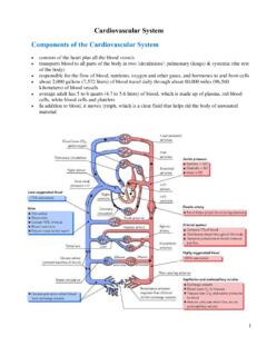
Cardiovascular System Components of the Cardiovascular …
www.soinc.org3 • Right Atrium: It collects deoxygenated blood returning from the body (through the vena cava) and then forces it into the right ventricle through the tricuspid valve. • Left Atrium: It collects oxygenated blood returning from the lungs and then forces it into the left ventricle through the mitral valve. • The atrioventricular (AV) valves (Mitral & Tricuspid Valves) prevent flow from the

Cardiovascular Pathology - Lecturio
cdn.lecturio.comCauses of a pathological S3 include conditions that reduce left ventricular compliance, such as left ventricular failure, left ventricular dilation, aortic regurgita-tion, mitral regurgitation, patent ductus arteriosus, and a ventricular septal defect. Conditions with reduced right ventricular compliance can also cause a pathological S3.
Reptile Cardiology: A Review of Anatomy and Physiology ...
vetmed.illinois.eduthe right to left to ensure that blood flow contin-ues to the systemic circulation. Once normal breathing resumes, pulmonary resistance de-creases, the HR increases, and the shunting of the blood is discontinued. These physiologic changes may occur when certain anesthetics are used. Dis-sociative agents, alpha-2 agonsists, and propofol
Left Ventricular Assist Device - AnMed Health
anmedhealth.orgLeft Ventricular Assist Device – Design •Design - Valve-less - Only one moving part (rotor) - Rotor spins on blood-lubricated bearings (rubies) designed for minimization of blood damage - All motor drive and control electronics are outside of the implanted blood pump •Speed range: 6,000 to 15,000 rpm •Flow range: 3 – 10 L/min

Pacemaker Learning Package - Agency for Clinical Innovation
www.aci.health.nsw.gov.au1] Tricuspid valve: separates the Right atrium from the Right ventricle 2] Bicuspid valve [Mitral valve]: lies between the Left atrium and Left ventricle. • Semi lunar Valves: 1] Pulmonic valve: is the valve between the Right ventricle and the pulmonary artery. 2] Aortic valve: is the valve between the Left ventricle and the aorta.
Electrocardiograms Made Easy! Part I. Basic ECG ...
www.elearnonline.netcontraction before ventricular involvement. After a small delay the impulse travels from the AV node to the ventricles through another specialized highway located in the septum of the ventricles. This highway is called the Bundle of His. The Bundle of His branches into the left and right bundle branches, and each delivers the