Transcription of Vital Pulpotomy vs - Hale Veterinary Clinic
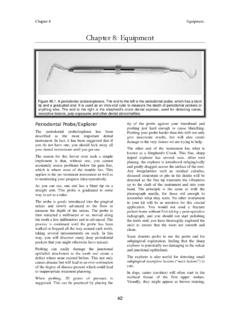
1 ,DVM,FAVD,DiplAVDCPage1 March,2000 LongDistance:1-866-866-8483 I have mentioned several times in past issues of The CUSP, broken teeth are a serious problem that definitely require treatment of some sort. In this item, I will assume that you understand that and I am going to concentrate on treatment planning for these fractured teeth. For now, let us consider the simplest of cases in which a dog has taken the tip off a canine tooth, The pulp is exposed, but there is no damage near or below the gumline and the remaining two thirds of the crown is intact. In cases where the pulp is exposed, there are two main treatment options to consider.
2 The first is extraction. This would achieve the objectives of removing a source of considerable pain as well as a conduit of infection. The other option is endodontic treatment of some type. Endodontic treatment options include total pulpectomy or partial Vital Pulpotomy . Each option has its advantages and disadvantages, indications and contra-indications. To understand some of these, it is important to know something of dental development and physiology When a permanent tooth is developing within the jaw of a young animal, it is constructed from the outside-in.
3 That is to say, the enamel of the crown is produced early in the process so that the outside dimension of the crown is established early. Once the enamel is formed, the tissue that made it goes dormant and no more enamel can ever be produced for that tooth. On the inside of the tooth is the pulp (blood vessels, nerves, lymphatics and various free cellular elements). Lining the inside wall of the developing tooth is a single layer of low columnar cells known as odontoblasts. These cells produce dentin, which is the hard tissue that makes up the bulk of the tooth. During pre-eruptive development and during eruption, the odontoblasts produce primary dentin.
4 Once the tooth had developed to its final length, the odontoblasts produce secondary dentin such that the pulp chamber inside the tooth gets smaller as the wall of the tooth gets thicker. This progression can be seen in the series of radiographs, all of the same animal and taken at 6, 14 and 32 months of age. In the first radiograph (six months of age), the tooth is still erupting, the dentin wall of the crown and root is very thin and the apex (root tip) has not yet formed. At 14 months, the tooth is fully erupted, the apex has closed and the dentin walls in the root and crown are much thicker than at six months.
5 By 32 months, the pulp chamber is smaller still and the root wall thick enough for these teeth to be considered mature . As long as the pulp remains healthy, the odontoblasts will continue to produce secondary dentin and the pulp chamber will get progressively narrower as the animal ages. This process occurs in most carnivores and omnivores. ,DVM,FAVD,DiplAVDCPage2 March,2000 LongDistance:1-866-866-8483 In dogs and cats, after the apex is closed , there are still several tiny channels through which the pulp communicated with the rest of the body.
6 This collection of channels constitutes the apical delta. Another point to keep in mind is that dentin is elastic. Due to a higher collagen and water content than enamel, it is not as hard, but neither is it as brittle. Therefore, a living tooth can bend a little without fracturing. This elasticity is thought to depend on a continuing source of moisture as supplied by the Vital pulp . With that understood, let s look at Vital Pulpotomy and total pulpectomy. In Vital Pulpotomy , also known as partial Vital pulpectomy, only the coronal portion of the pulp is removed. Typically, a sterile bur, in a high-speed hand piece is used to amputate the pulp to a depth of about 6-8 millimeters.
7 The rest of the pulp is left inside the tooth. Next, various dental materials are placed inside the chamber on top of the pulp stump to protect the pulp and to seal the tooth against the ingress of bacteria that would kill the pulp . Typically, the material placed in direct contact with the freshly cut pulp stump is a calcium hydroxide preparation. The very high pH actually causes a surface necrosis of the pulp it touches. Undifferentiated free cells in the pulp below the zone of necrosis are stimulated to become odontoblasts and start producing dentin (this dentin produced in response to some irritation is known as tertiary or reparative dentin).
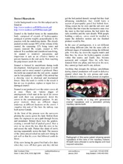
8 The bulk of the pulp inside the tooth remains unaffected by the treatment and so normal secondary dentin production continues as before within the rest of the tooth. This radiograph shows the dentin bridge that has formed between the Vital pulp and the restorative materials placed inside the pulp chamber of the crown of this canine tooth. As the animal ages, the chamber in the root should get smaller due to secondary dentin production. In total pulpectomy, the entire pulp is removed from the tooth and the chamber (root canal) is filled with dental materials. Once this is done, the odontoblasts are all gone there can be no more dentin production of any kind in the treated tooth.
9 Its wall will never get any thicker no matter how long the animal lives. This radiograph of a cat mandible shows the mandibular canines after the canals have been debrided and obturated (filled), but prior to final restoration of the access holes in the crowns. The advantages to partial Vital pulpectomy and direct pulp capping include: usually faster and cheaper than total pulpectomy, keeps the pulp intact so the tooth can continue to mature, maintains a source of moisture to the dentin, helping to keep it elastic (flexible). The big disadvantage is, it may not work! If a partial Vital Pulpotomy and direct pulp capping is done and it fails, that means that the pulp dies anyway.
10 During the time the pulp is dying, there will be acute toothache. Once the pulp is dead, you have the same problems as you get with an untreated broken tooth chronic pain and infection. In fact, with the crown sealed with a bonded restoration, any pressure that builds up inside the tooth (due to gas production by bacteria) must be released through the apical delta. To monitor for signs of success or failure, it is very important that the tooth be re-radiographed (intra-oral dental radiographs under general anesthesia) at least once (six to twelve months post-operatively) and preferably every few years thereafter.