Transcription of Examination of the Peripheral Blood Smear
1 Examination of the Peripheral Blood 1 of 7 Printed 1/14/13 Created Lisa Senzel Asst. Prof. - Clinical Path 5/9/2008 Revised Bruce Kube Day Heme Tech 1 10/23/2012 Reviewed Jay Bock 4/10/2012 Approved Lisa Senzel Asst. Prof. - Clinical Path 1/10/2013 Replaces 5/08 HEM Procedure: Examination of the Peripheral Blood Smear (How to Perform) I. PRINCIPLE The Examination of the Peripheral Blood Smear is an important basic hematological procedure. Many hematological diagnoses depend upon this procedure and often a definitive diagnosis can be established from the careful Examination of the Blood film. The Smear is stained with Wright s stain and performed after the complete Blood count is run. A manual differential consists of: 1. The WBC differential count with visual confirmation of WBC count 2. RBC morphology 3.
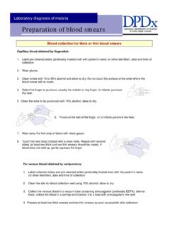
2 Estimation of the platelet count II. SPECIMEN Whole Blood collected in a lavender Vacutainer tube or lavender Microtainer containing K2 EDTA are used. These collection devices must be properly collected and filled as per manufactures recommendations. The sample must be free of clots and hemolysis. A. Sample Collection Samples are collected as per phlebotomy protocols. B. Sample Preparation The Smear will be prepared either automatically by the LH Slidemaker, or manually, in the case of microtainers and samples that are not run in the primary mode on the Coulter and the ACP lab. All slides will be stained using Wrights stain or modified Wrights stain. Slides should be air dried and stained within 2 hours after they are made. III. REAGENTS Examination of the Peripheral Blood 2 of 7 Printed 1/14/13 Wrights stain is used and is prepared per the procedure in this manual.
3 Modified Wright stain is purchased in prefilled stain packs IV. PROCEDURE When performing a Blood film Examination the entire slide is to be evaluated regardless of the reason that the evaluation was initiated. All slides are reviewed for RBC, WBC and platelet anomalies and all significant findings are to be reported. 1. Scanning the Smear : Use low power (10X) Objective a. Check quality of Smear and stain b. Determine distribution of cells: 1. Check for large platelet clumps 2. Presence of rouleaux formation of RBCs 3. Check for WBC clumping c. Select best area for detailed morphological evaluation: 1. Edges of erythrocytes not quite touching each other 2. Devoid of broken areas 3. No flattening or distortion of RBCs usually seen at the extreme periphery of the Blood Smear 2. WBC Estimation: Use (40X) High Dry Objective 1.
4 To estimate total WBC count/mm 3. Count the number of both intact and disruptive WBCs in each of 10 microscopic fields in different areas of the slide where RBCs slightly overlap. Divide the total number by 10 to establish the mean number of WBC/field and multiply this mean by 2,000 to get the estimated WBC. WBC per HPF Estimated WBC 2 - 4 4,000 7,000 4 - 6 7,000 - 10,000 6 - 10 10,000 - 13,000 10 - 20 13,000 - 18,000 3. White Cell Differential: Use (50X) Oil Emersion Objective 1. Count and identify 100 WBCs and record them on the computer keyboard as per LIS procedure. If any nucleated RBCs are seen, count them separately, they are not to be included in the 100 cell count. While counting WBCs make note of any inclusions or abnormalities in the WBCs (ex.)
5 Dohle bodies, toxic granulation, vacuoles). 2. WBCs should be read in an area where RBCs are not quite touching each other. It is acceptable to move into a slightly thicker area, but once there are several RBCs touching each other (ie, 4 RBCs touching), and/or the stain becomes darker due to thickness, the area is considered unreadable. If there are fewer than 50 WBCs in Examination of the Peripheral Blood 3 of 7 Printed 1/14/13 the readable area, release the automated differential with one of the following comments: WCOM1: An accurate manual diff can not be performed, too few cells in readable area, no definite blasts. WCOM2: An accurate manual diff can not be performed, too few cells in readable area, suspicious for blasts, however blasts cannot be enumerated by automated hematology analyzer. 4. RBC/WBC Morphology and Platelet Estimation: Use (100X) Oil Emersion Objective 1.
6 Scan 10 microscopic fields (approximately 500 RBC/OE field) in different areas of the Smear with evenly dispersed RBCs for the erythrocyte evaluation morphology. Note any variations from normal and classify them as 1+, 2+ or 3+. Examine for size, shape, relative hemoglobin content, polychromatophilia, inclusions and rouleaux formation Etc. See chart below. 2. Perform platelet estimation and morphology (ex. large , giant). The detailed procedure for platelet estimation is found in this manual (HEM ). Examination of the Peripheral Blood 4 of 7 Printed 1/14/13 Standardization of RBC morphology reporting In order to standardize RBC morphology reporting, the following conventions have been adopted. RBC Morphology Grading** Finding 1+ 2+ 3+ Notes Macrocytosis 6-15 16-30 >30 Approximate # of large RBC 95 - 108fl 109 - 120fl >120fl MCV Microcytosis 6-15 16-30 >30 Approximate # of small RBC 76-80 66-75 <65 MCV Hypochromia** 6-15 16-30 >30 Avg # of Hypochromic cells /10 fields.
7 (>1/3 central palor) < MCHC Anisocytosis 6-15 15-30 >30 Avg of microcytic and macrocytic cells /10 fields > RDW Poikilocytosis ( 2 abnormal shapes) 2-5 6-15 >15 Sum of avg # of all types of abnormally shaped RBC /10 fields Teardrop Cells 2-5 6-15 >15 Avg # seen/10 fields Target cells 2-5 6-15 >15 Avg # seen/10 fields Ovalocytes 2-5 6-15 >15 Avg # seen/10 fields Stomatocytes 2-5 6-15 >15 Avg # seen/10 fields Schistocytes 2-5 6-15 >15 Avg # seen/10 fields Spherocytes 1-5 6-15 >15 Avg # seen/10 fields Polychromasia 2-4 5-7 >7 Avg # of Polychromatophilic cells /10 fields. Sickle Cells 1-5 6-15 >15 Avg # of Sickle cells/10 fields Burr Cells 2-5 6-15 >15 Avg # seen/10 fields Rouleaux 1-5 6-15 >15 Avg # Rouleaux forms seen/10 fields RBC inclusions Howell-Jolly Bodies Pappenheimer Bodies Basophilic stippling Cabot rings 1-2 3-5 >6 Avg # of inclusions in RBC /10 fields.
8 ** All values are in approximate # based on 100x OEF of approximately 100-150 RBC/field **When evaluating the MCHC, all samples MCHCs > should be checked for hemolysis, agglutination or insufficient sample volume To obtain average, count the number of cells of interest in each of 10 fields and divide by 10 Examination of the Peripheral Blood 5 of 7 Printed 1/14/13 Finding** Toxic Granulation Present Toxic Vacuolation Present Present D hle bodies Present Present Hypersegmentation Present Present Present Pelger-Huet/Pseudo Pelger-Huet (Hyposegmentation) Present Hypogranularity Present Smudge cells ** Present ** Above morphological characteristics are to be indicated as present if noted on Smear .
9 Platelet Morphology** Giant Platelets Present Large Plt Present Agranular (gray) Plt Present Platelet Satellitosis Present ** Above morphological characteristics are to be indicated as present if noted on Smear . QUALITY CONTROL Blood smears prepared in the hematology lab will be retained for a period of not less than one year. Slides will be filed by date and then by accession number within the date. In the event of a downtime, slides prepared from specimens accessioned during downtime will be filed at the beginning of the days work according to the first character of the downtime accession number (Z or C). In the event a sample result is not in need of Peripheral Smear review (does not fail any review criteria), all results will be archived to diskette and kept for a period of 6 months. These results serve as the Smear in this case.
10 All abnormal scatterplots will be kept for a period of not less than three months. The ACP lab will make and retain Blood smears on all patients regardless of Smear review, for a period of not less than one year. Interpretation of Results When studying a stained Smear do not go too far into the thick area of the slide. The morphological characteristics of the cells are difficult to distinguish in this area. Also, do not use the thin edge of the Smear where the RBCs appear completely filled with hemoglobin and show no central pallor. The cells in this area are generally distorted and will not show a true morphological picture. Examination of the Peripheral Blood 6 of 7 Printed 1/14/13 When the WBC is below 1000/cumm, it will be difficult to find many white cells on the stained Smear . The differential can be done by counting fewer cells.