Search results with tag "Microscopy"

Measurement of Fibers
www.cdc.gov5 Electron microscopy FI-17 6 Scanning electron microscopy (SEM) FI-18 7 Transmission electron microscopy (TEM) FI-18 8 Optical detection (light scattering) FI-20 9 Fiber classification FI-21 10 Conclusions FI-22 11 References FI-23. NIOSH Manual of Analytical Methods 5th Edition Chapter FI April 2016 Page FI-2 of FI-31 ...

MICROENCAPSULATION - Jiwaji University
www.jiwaji.eduConventional light microscopy-Used to determine the shape and outer structure of the microparticles. Scanning electron microscopy-It can be used for the investigation of double walled systems. Conflocal fluorescence microscopy-35 used for the structure characterization of multiple walled microspheres.
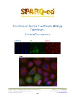
Introduction to Cell & Molecular Biology Techniques ...
di.uq.edu.auTransmission electron microscopy provides a cross section of a specimen, while scanning electron microscopy gives a three-dimensional image of the surface of a specimen. Fluorescence Microscopy – uses fluorescent materials to indicate structures in a specimen.

DIAGNOSTICS, MEDICAL DEVICES & OTHER HEALTH …
stoptb.orgGDF SMEAR MICROSCOPY KITS This kit provides a convenient way to receive all of the needed reagents and consumables to perform 1,000 smears using bright field (ZN) microscopy. Contents: DIAGNOSTIC CONSUMABLES KIT – ZN LIGHT MICROSCOPY Product Code: 106522 This kit provides a convenient way to receive all of the needed

Chapter 4 –Microscopy - Quantitative Light Imaging ...
light.ece.illinois.edu4.5 PhaseContrast Microscopy ECE 460 –Optical Imaging Note: For a = 0 recover Dark Field Microscopy Assume“small”phaseshift Assume small phase shift cos 1; 2 2 2 2 (, ) 2 sin sin 2(,)sin Ixy a a aaxy PC couples into intensity

Transmission Electron Microscopy -TEM- - Jiwaji University
www.jiwaji.eduTransmission Electron Microscopy-TEM-The first electron microscope was built 1932 by the German physicist Ernst Ruska, who was awarded the Nobel Prize in 1986 for its invention. He knew that electrons possess a wave aspect, so he believed he could treat them in a fashion similar to light waves. Ruska

Synthesis of Graphene Oxide (GO) by Modified Hummers ...
file.scirp.orgmicroscopy (SEM), high-resolution transmission electron microscopy (HR-TEM), Fourier transform infrared spectroscopy (FTIR), ultraviolet-visible spectroscopy, Raman spectroscopy, Brunauer-Emmett-Teller (BET) surface area analysis and dif-ferential scanning calorimetry (DSC) and thermogravimetric analysis (TGA).

ASBESTOS and OTHER FIBERS by PCM 7400
www.cdc.govThis method can be used in conjunction with electron microscopy (e.g., Method 7402) for assistance in identification of fibers. For fibers with diameters >1 μm, polarizing light microscopy (as in NIOSH Method 7403) may be used to identify …

Basic Hematology
www.hematology.orgblood smear is examined carefully using 40 x to 100 x objective – usually oil immersion lens 2. 100 white blood cells are counted 3. Cells are classified by ... it is determined by light microscopy. d) it is determined by electron microscopy. e) it is derived from the white cell count.

Research proposal - University of Groningen
www.rug.nlStudy of the films by AFM, far and near field optical microscopy, electron microscopy and X-Ray diffraction techniques September 2011–August 2012 Specroscopic study of the samples using time resolved spectroscopy, optical pump-probe and transient absorption techniques Study of the charge transport properties of

Medical Microbiology, Virology & Immunology
elib.vsmu.bySTED-microscopy – stimulated emission-depletion fluorescent microscopy . STX toxin – Shiga toxin . T3SS – type III secretion system . T4SS – type IV secretion system . T7SS – type VII secretion system . TBEV – tick-borne encephalitis virus . Tc – T cytotoxic cell . TCA – tricarboxylic acid . TCBS agar – thiosulfate-citrate ...
Materials: Structure, Properties, and Performance
assets.cambridge.orgScanning probe microscopy Auger electron spectroscopy Transmission electron microscopy Creep Fatigue Strength Toughness Dynamic response Constitutive response Characterization Mechanical Properties Processing Continuum mechanics Theory Computational mechanics Quantum mechanics Crystallography, defects
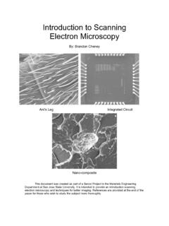
Introduction to Scanning Electron Microscopy
www.sjsu.eduFeb 01, 2005 · In scanning electron microscopy visual inspection of the surface of a material utilizes signals of two types, secondary and backscattered electrons. Secondary and backscattered electrons are constantly being produced from the surface of the specimen while under the electron beam however they are a result of two separate types of interaction.
如何展开药物多晶型研究?
www.crystalpharmatech.com.cnAll variants of spectroscopy, microscopy, scattering etc. are used Structure, order and dynamics of the solid state probed Spectroscopy Microscopy Scattering Thermal analysis Physio-Mechanical analysis Computer Modeling ssNMR, IR, Raman, Terahertz… SEM, Optical, AFM… X-ray, Laser, … DSC, TGA, Microcalorimetry, DMA, DEA…

Atomic Force Microscopy (AFM)
users.metu.edu.trAtomic Force Microscopy (AFM) 1. General Principle The Atomic Force Microscope is a kind of scanning probe microscope in which a topographical image of the sample surface can be achieved based on the interactions between a tip and a sample surface. The atomic force microscope was invented by Gerd Binning et al. in 1986 at IBM Zurich based on ...

AFB Smear Microscopy - APHL
www.aphl.orgSputum Smear Results •In 2010, 43% of pulmonary TB cases in the U.S. were sputum smear positive •Incremental diagnostic yield of examination of three sputum specimens among smear positive cases First specimen Second specimen Third specimen 85.8% 11.9% 3.1% CDC. Reported Tuberculosis in the United States, 2010.

Lipophilic Tracers—Dil, DiO, DiD, DiA, and DiR
tools.thermofisher.commoderate fluorescence quantum yields, and short excited state lifetimes in lipid environments (~1 ns).34 They are insoluble in water, but their fluorescence is readily detected when incorpo-rated into membranes. A summary of spectral properties is shown in Table 2, together with appr o priate filter sets for fluorescence microscopy applications

Practical issues in FTIR spectroscopy Lab/demo section ...
warwick.ac.ukFlexible sampling - any phase or size - no preparation 1µm sample area - Raman microscopy possible Glass cells - good medium for cell design, low cost Fiber optics - up to 100m, routine Water - weak scatterer - excellent solvent Enhanced by resonance, surface interactions
Official American Thoracic Society/Infectious Diseases ...
www.thoracic.orgcopy be performed, rather than no AFB smear microscopy, in all patients suspected of having pulmonary TB (strong recommendation, moderate-quality evidence). Remarks: False-negative results are sufficiently common that a negative AFB smear result does not exclude pulmonary -mon that a positive AFB smear result does not confirm pulmonary TB.
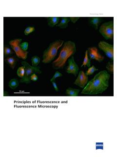
Principles of Fluorescence and Fluorescence Microscopy
pages.zeiss.comImportantly, a major breakthrough for the use of small or - ganic dyes as fluorescent labels came in 1941, when they were first conjugated to antibodies, thereby inaugurating the field of immunofluorescence5. Direct fluorescence Indirect fluorescence Fluorophore Primary antibody Secondary antibody Antigen

Scanning Electron Microscopy Working Principle
assets.thermofisher.comAn electron source—also referred to as an electron gun—emits electrons that get accelerated by an applied voltage. Magnetic lenses converge the stream of electrons into a focused beam, which then hits the sample surface in a fine, precise spot. The electron beam then scans the surface of the specimen in a rectangular raster.
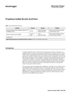
Propidium Iodide Nucleic Acid Stain
assets.thermofisher.comrescence microscopy, centrifuge the sample, remove the supernatant, and resuspend the cells in fresh buffer. Apply a drop of the suspension to a microscope slide, cover with a coverslip, and view using appropriate filters (see Fluorescence Spectral Characteristics). Counterstaining the Specimen by Chromosome FISH Preparing the Sample
Official American Thoracic Society/Infectious Diseases ...
www.cdc.gov• We recommend that acid-fast bacilli (AFB) smear micros-copy be performed, rather than no AFB smear microscopy, in all patients suspected of having pulmonary TB (strong recommendation, moderate-quality evidence). Remarks: False-negative results are sufficiently common that a negative AFB smear result does not exclude pulmonary TB.

Diagnosis of Tuberculosis Disease - Centers for Disease ...
www.cdc.govThe presence of acid-fast-bacilli (AFB) on a . sputum smear. or other specimen often indicates TB disease. Acid-fast microscopy is easy and quick, but it does not confirm a diagnosis of TB because some acid-fast-bacilli are not . M. tuberculosis. Therefore, a . culture. is done on all initial samples to confirm the diagnosis. (However, a ...
MGITTM Procedure Manual - FIND
www.finddx.orgMicroscopy 4. Reporting . MGITTM Procedure Manual 4 ... The introduction of the BACTEC 460 TB System revolutionized laboratory ... resulting in fluorescence within the MGIT tube when visualized under UV light. The intensity of fluorescence is directly proportional to the

Tuberculosis in England
assets.publishing.service.gov.ukof people notified did not have any laboratory results reported (culture, microscopy, PCR, or histology) to confirm their TB diagnosis. As in previous years, culture confirmation was higher for pulmonary (75.3% of cases) than non-pulmonary cases (44.2%). Culture confirmation rates in children are much lower at 27% for pulmonary disease, and 32.7%

DETECTION OF GRAPHENE IN COVID19 VACCINES
filedn.commicroscopy allows the excitation laser to be focused on specific objects and points ... the effects of matrix fluorescence. The spectra were analyzed with SPECTRA MANAGER software, version 2. JASCO ... (introduction of functional groups) of the network
Optical Microscope; • Scanning Electron Microscope (SEM ...
my.eng.utah.eduprototype electron microscope in 1931, capable of four-hundred-power magnification; the apparatus was the first demonstration of the principles of electron microscopy. Two years later, in 1933, Ruska built an electron microscope that exceeded the resolution attainable with an optical (light) microscope.

Hematology and Clinical Microscopy Glossary
documents.cap.orgBlood Cell Identification The segmented neutrophil is the predominant blood leukocyte. It has a similar size to a band neutrophil (ie, 10 to 15 μm in diameter), as well as comparable shape (round to oval), and cytoplasmic appearance (pale pink cytoplasm with specific granules). The N:C ratio is 1:3 and the nuclear chromatin is highly condensed.

A New Tool to Diagnose Tuberculosis:The Xpert MTB/RIF Assay
www.cdc.govradiographic, and other laboratory findings. The Xpert MTB/RIF assay does not replace the need for smear with microscopy for acid-fast bacilli, culture for mycobacteria, and growth-based drug susceptibility testing, in addition to genotyping for early discovery of outbreaks.

T Cell TransAct™ human - Miltenyi Biotec
www.miltenyibiotec.com2.1 Sample preparation ... by flow cytometry or fluorescence microscopy. nless therwse spec⁶call ncate, all lten tec pructs an serces page 3/3 140-004-952.05 are r research use nl an nt r agnstc r therapeutc use. 3. Refer to Examples of T cell activation and expansion ... fluorescence. Negative control

Leica TCS SP8
www.leica-microsystems.comof fluorescence spectra ... of older detection principles, such as pixel convolution, noise, and voltage-related scaling issues. backed by lightning-fast counting electronics, even ... microscopy has an inherent limitation of serial image collection. Camera-based

Virology Techniques - Texas A&M AgriLife
agrilife.orgor fluorescence, designed to identify the virus of inter-est are used. This label then enables the visualization of the virus cluster (because a single virus is too small to see with a light microscope) with the light micro-scope, in the case of peroxidase, or an ultraviolet (UV) light microscope in the case of fluorescence. Electron Microscopy

State Program Management Unit
upnrhm.gov.inoptions for filling online applications. (1) eligibility criteria and registration: s. no program name sub program position age ... smear microscopy. 1. candidates with higher qualification ( for example graduates ) shall be ... bacteriology (or) 2. five years of work experience in tb ...

NATIONAL TUBERCULOSIS CONTROL PROGRAM MANUAL …
doh.gov.phSection 2.2. Diagnosis of tuberculosis disease 18 DEFINITION OF TERMS 18 POLICIES 19 PROCEDURES20 A. Collection and transport of sputum specimens 20 B. Procedure for Xpert MTB/RIF 22 C. Procedure for smear microscopy 22 D. Decision on diagnosis based on laboratory results 23 E. Decision on further testing based on result of Xpert MTB/RIF 26

Basic Image Processing with FIJI/ImageJ
www.jmu.edumicroscopy facility director. • Except for ethically applied simple linear histogram (intensity) adjustments, reducing bit depth, and cropping, ALL image processing procedures MUST be disclosed in your Methods section and/or figure legend. 1.2 CONVENTIONS USED IN …

What Procedures are Performed by Primary Care Providers?
2016forum.paeaonline.orgOct 17, 2014 · Microscopy (Urinalysis, KOH, Gram Stain, Wet Mount) Dermatology Procedures Dermatologic Procedures (acne surgery, biopsies, electrosurgery, cryosurgery, chemosurgery, surgical curettement) Incision and Drainage Nail Excision Simple Wound Closure Complex Wound Closure Wound Care GI Procedures

*134315810* - Dr Lal PathLabs
www.lalpathlabs.com(Microscopy) Note: A Single negative smear does not rule out malaria Dr. Anil Arora MD (Pathology) Consultant Pathologist Dr Biswadip Hazarika MD (Pathology) Consultant Pathologist Dr. Shalabh Malik MD (Microbiology) HOD Micro & Clinical Path-----End of report ----- PatientReportSCSuperPanel.GENERAL_METHOD_SC ...

Mycobacterial Culture Final - APHL
www.aphl.org– ~10 viable bacilli/ml of sputum needed for culture compared to at least 5000 bacilli/ml of sputum for microscopy • Culture used for species identification, drug susceptibility testing (DST), and genotyping • Culture also used to monitor patient response to treatment 3

leyenda marca de agua - IDENTIFICATION OF POSSIBLE …
www.laquintacolumna.infopia_Micro-RAMAN Correspondingly, we have highlighted that graphene has a multiplier effect on the radiation ... After wide intervals of observation under optical microscopy, using different light filters and magnification qualities, objects compatible with the appearance of graphene have been

A2018ReferenceGuidetotheBanffClassification of Renal ...
banfffoundation.orgmission electron microscopy (EM). Depending on clinical and histopathological findings a complete nephropathological work-up including staining for immunoglobulin heavy and light chains and complement split productsmight be necessary to rule out or confirm a di-agnosisofglomerulonephritis.Otherancillarystainingmight

Microscopy I Light and Electron Microscopy
www.auburn.eduLight Microscopy Bright field Microscopes--the most common general use microscopes. Bright field microscopes are named because the microscopic “field” is bright, while the object being viewed is dark. - Simple design - Light directed at specimen is absorbed to form image

MICROSCOPY I. OBJECTIVES II. INTRODUCTION
www.sas.upenn.eduII. INTRODUCTION There are several types of microscope (simple, compound, light or bright-field, dark-field, electron, fluorescence, interference, etc.) but the one most commonly used for bacteriological purposes is the bright-field or light microscope. This microscope is
Similar queries
Electron microscopy, Transmission Electron Microscopy, MICROSCOPY, Fluorescence microscopy, SMEAR MICROSCOPY, Light, RAMAN, Analysis, Light Microscopy, Basic Hematology, Smear, Research proposal, Study, Using, Structure, Scanning, Electron, Introduction to Scanning Electron Microscopy, Scanning electron microscopy, Atomic Force Microscopy AFM, Principle, AFB smear microscopy, Sputum, TUBERCULOSIS, Fluorescence, Preparation, Sample, American Thoracic Society, AFB smear, Principles of Fluorescence and Fluorescence Microscopy, Breakthrough, Scanning Electron Microscopy Working Principle, Smear micros-copy, Procedure Manual, INTRODUCTION, Optical Microscope; • Scanning Electron Microscope, Clinical Microscopy Glossary, Blood Cell Identification, Blood, Laboratory, Sample preparation, Of fluorescence, Principles, Virology Techniques, Applications, Bacteriology, Diagnosis of tuberculosis, Diagnosis, Procedures, What Procedures are Performed by Primary Care Providers, Clinical