Search results with tag "Fluorescence"

SPECTROMETRIE DE FLUORESCENCE MOLECULAIRE
univ.ency-education.comSPECTROMETRIE DE FLUORESCENCE MOLECULAIRE La spectrométrie de fluorescence moléculaire étudie l’émission de lumière par des molécules, en solution après excitation par des photons appartenant au domaine du visible ou du proche ultraviolet. PARTIE I : PHENOMENE DE FLUORESCENCE I. TERMINOLOGIE : 1.
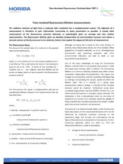
Time resolved fluorescence lifetime measurements
www.horiba.comThe extra specificity of the fluorescence lifetime allows easy discrimination against scattered excitation and background fluorescence. Determination of Förster resonance energy transfer (FRET) is much simpler using the fluorescence lifetime, …

14. Total Internal Reflection and Evanescent Waves
www.brown.edufluorescence (TIRF) microscopy Only objects within ~100 nm of the interface are illuminated. conventional fluorescence microscope image is blurred due to fluorescence from out-of-focus sources TIRF image is sharper

Les principes de la spectroscopie de fluorescence : de la ...
www.heliospir.netRendement quantique: efficacité relative de la fluorescence comparée aux autres voies de désexcitation F = nombre de photons émis Grandeur sans dimension 0,05 à 1 nombre de photons absorbés F = 0,85 pour la fluorescéine Intensité de fluorescence : dépend de la longueur d’onde d’excitation ex alors que l’allure du
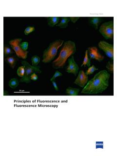
Principles of Fluorescence and Fluorescence Microscopy
pages.zeiss.comImportantly, a major breakthrough for the use of small or - ganic dyes as fluorescent labels came in 1941, when they were first conjugated to antibodies, thereby inaugurating the field of immunofluorescence5. Direct fluorescence Indirect fluorescence Fluorophore Primary antibody Secondary antibody Antigen
Real-Time PCR Applications Guide - Bio-Rad Laboratories
www.bio-rad.comInitially, fluorescence remains at background levels, and increases in fluorescence are not detectable (cycles 1–18 in Figure 1.1) even though product accumulates exponentially. Eventually, enough amplified product accumulates to yield a detectable fluorescent signal. The cycle number at which this occurs is called the threshold cycle, or C T ...

Virology Techniques - Texas A&M AgriLife
agrilife.orgor fluorescence, designed to identify the virus of inter-est are used. This label then enables the visualization of the virus cluster (because a single virus is too small to see with a light microscope) with the light micro-scope, in the case of peroxidase, or an ultraviolet (UV) light microscope in the case of fluorescence. Electron Microscopy

1 Basic Principles of Fluorescence Spectroscopy
application.wiley-vch.de1 Basic Principles of Fluorescence Spectroscopy 1.1 Absorption and Emission of Light As fluorophores play the central role in fluorescence spectroscopy and imaging we
MGITTM Procedure Manual - FIND
www.finddx.orgMicroscopy 4. Reporting . MGITTM Procedure Manual 4 ... The introduction of the BACTEC 460 TB System revolutionized laboratory ... resulting in fluorescence within the MGIT tube when visualized under UV light. The intensity of fluorescence is directly proportional to the

AFB Smear Microscopy - APHL
www.aphl.orgFluorescence Microscopy • A fluorescence microscope is required for examining fluorochrome-stained smears: –Mercury vapor or halogen bulb light source (about 150 hours of use) –Newer mercury bulbs (about 2,000 hours of use) –LED Bulbs (about 15,000 hours of use) –Excitation and emission (barrier) filters are necessary for
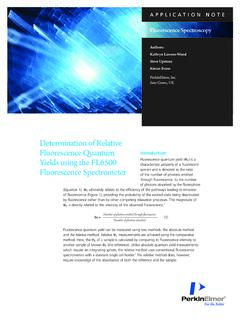
Determination of Relative Fluorescence Quantum Yields ...
resources.perkinelmer.com0.01 – 0.1 at the excitation wavelength of 537 nm. The absorbance of each solution was determined using a PerkinElmer LAMBDA 265 UV/Vis Spectrophotometer using PerkinElmer UV Lab™ software. In the absence of a separate UV-Vis instrument, the PerkinElmer FL6500 or FL8500 Fluorescence Spectrometer’s

jpg->pdf - ilovepdf
www.scei-concours.frET J.DE BAERDEMAEKER : The use of front face fluorescence spectroscopy to classify the botanical origin of honey samples prod uced in Switzerland : Elsevier, 2007, p.314-323 DOT Ill Rencontre lors d'animations culturelles pendant l'été 2017 avec un physicien ayant découvert par hasard la fluorescence de l'acacia.

INTRODUCTION TO MEDICAL AND MOLECULAR BIOLOGY
www.fmed.uniba.sk1.2.3 Fluorescence microscope Fluorescence microscope belongs to light microscopes which use vapour lamp as a source of UV radiation. Specially prepared biological samples are illuminated with light of a specific wavelength which induces emission of light with longer wavelength – visible light.

T Cell TransAct™ human - Miltenyi Biotec
www.miltenyibiotec.com2.1 Sample preparation ... by flow cytometry or fluorescence microscopy. nless therwse spec⁶call ncate, all lten tec pructs an serces page 3/3 140-004-952.05 are r research use nl an nt r agnstc r therapeutc use. 3. Refer to Examples of T cell activation and expansion ... fluorescence. Negative control
Propidium Iodide Nucleic Acid Stain
assets.thermofisher.comApproximate fluorescence excitation/emission maxima: 535/617 nm, bound to nucleic acids Propidium Iodide Nucleic Acid Stain Introduction Propidium iodide (PI) binds to DNA by intercalating between the bases with little or no sequence preference and with a stoichiometry of one dye per 4–5 base pairs of DNA.1 PI also
Chapter 6 Photoluminescence Spectroscopy
ocw.utm.myIt emits energy from an excited electronic state as light. Some of the incident energy is absorbed and re-emitted as light of a longer wavelength (Stoke’s law). ... Measure distances using molecular rulers: fluorescence resonance energy transfer (FRET) Band gap of semiconductors Nanomaterials characterization .

dots for two-photon fluorescence imaging SUPPORTING ...
www.rsc.orgrecorded with a tapping mode in air using Multimode 8 atomic force microscope (VEECO, USA). X-ray diffraction (XRD) pattern was characterized on D8 advance X-ray powder diffractometer (Bruker, Germany). Raman measurement was obtained by confocal Raman 35 microscope (CRM200, WITec) with 50× objective using excitation laser line of 532 nm.

Lipophilic Tracers—Dil, DiO, DiD, DiA, and DiR
tools.thermofisher.comApproximate fl uorescence excitation/emission maxima: See Table 2. Lipophilic Tracers—Dil, DiO, DiD, DiA, and DiR | 2 staining is usually less intense than that of DiI, and occasionally fails completely in fixed tissues.6,11 DiD (D307, D7757) is an analog of DiI with markedly red-shifted fluorescence excitation
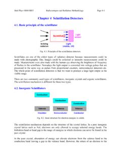
Chapter 4 Scintillation Detectors - McMaster Faculty of ...
www.science.mcmaster.caAll fluorescence emissions ... absorption of the photon and energy transfer to the electron in the photocathode material 2) the migration of the photoelectron to the surface of the photocathode 3) escape of the electron from the photocathode surface. Fig. 4.6. Basic elements of a PMT.

Scattering - Michigan Technological University
pages.mtu.eduscattered light is the same as the incident light (Rayleigh and Mie scattering) • Inelastic scattering – the emitted radiation has a wavelength different from that of the incident radiation (Raman scattering, fluorescence) • Quasi-elastic scattering – the wavelength (frequency) of the scattered light shifts (e.g., in moving matter due to

MICROENCAPSULATION - Jiwaji University
www.jiwaji.eduConventional light microscopy-Used to determine the shape and outer structure of the microparticles. Scanning electron microscopy-It can be used for the investigation of double walled systems. Conflocal fluorescence microscopy-35 used for the structure characterization of multiple walled microspheres.

MICROSCOPY I. OBJECTIVES II. INTRODUCTION
www.sas.upenn.eduII. INTRODUCTION There are several types of microscope (simple, compound, light or bright-field, dark-field, electron, fluorescence, interference, etc.) but the one most commonly used for bacteriological purposes is the bright-field or light microscope. This microscope is

Qubit 3.0 Fluorometer - Thermo Fisher Scientific
tools.thermofisher.comThe Qubit® 3.0 Fluorometer is a benchtop fluorometer that can be used for the quantitation of DNA, RNA, microRNA, and protein using the highly sensitive and accurate fluorescence-based Qubit® quantitation assays. Additionally, Ion Sphere™

DETECTION OF GRAPHENE IN COVID19 VACCINES
filedn.commicroscopy allows the excitation laser to be focused on specific objects and points ... the effects of matrix fluorescence. The spectra were analyzed with SPECTRA MANAGER software, version 2. JASCO ... (introduction of functional groups) of the network

Basic Image Processing with FIJI/ImageJ
www.jmu.edu• For a quick introduction to the layout of the ImageJ program, see here. • Navigation through menus and sub-menus is notated using >. For example, ^Image > Type _ tells you to go to access the Image menu and then the Type sub-menu. • True-color images are images of specimens with color (e.g. histological stains). Fluorescence

Fluorophore selection guide for flow cytometry - Inserm
cytobase.montp.inserm.fr→ Energy transfer dye excited at ~488 nm with emission maximum at ~670 nm → Also known as PE-Cy®5 → Bright fluorescence → Low compensation against RPE → Significant emission in APC channel with 633 nm excitation → Measured in FL3 channel on BD FACScan™ and BD FACSCalibur™ instruments
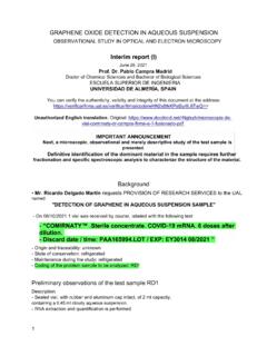
GRAPHENE OXIDE DETECTION IN AQUEOUS SUSPENSION
www.thecompleteguidetohealth.coma) Bright field. b-d) Fluorescence extinction microscopy (FQM) Kim et al, 2010. Seeing graphene-based sheets, Materials Today, Volume 13, 2010, Pages 28-38, Literature Image Low magnification TEM "The figure shows a TEM image of bilayer graphene with edges that tend to curl and bend slightly" 5

Module 1 (Molecular Spectroscopy) Lecture 1 Date
www.sctce.ac.inFluorescence spectroscopy, Phosphorescence spectroscopy . Beer Lamberts Law When a monochromatic light is passed through a dye solution, the absorbance is proportional to product of concentration and thickness of solution. A= c t [ is molar absorptivity, c is concentration and t is thickness of solution. ...

Production of X-rays - Western Kentucky University
www.wku.edufluorescence), filters with energy selective absorption edges are not used to harden the beam, but to obtain a more monochromatic beam (a beam with predominantly one energy). •By choosing the right element, it is possible to absorb a band of high energy photons preferentially over an adjacent band of low energy photons. Production of X-rays
FlowJo Basic Tutorial - flowlab-childrens-harvard.com
www.flowlab-childrens-harvard.comDrag to draw a gate to set the range of fluorescence values that will define FL1 positive cells. Name this subset FL1+. Click the plus sign to add the name to the list for future use. Click OK. There are several gating tools for bivariate plots. Rectangle, Polygon, Quadrant or Ellipse gating tools can be selected from the top of the Graph
Energy Dispersive X-ray Spectroscopy (EDS)
www.nuance.northwestern.eduEnergy Dispersive X-ray ... fluorescence •Sample prep –Flat, homogeneous, conductive, non-porous . Z Correction •Backscattering •Higher mean atomic number = higher backscatter yield (BSE do not general X-ray) •If mean atomic number of the sample is higher than standard,

MM12573 - Healthcare Common Procedure Coding System …
www.cms.govJan 20, 2022 · • 0243U - Time-resolved fluorescence immunoassay of placental-growth factor in maternal serum to evaluate risk of preeclampsia • 0244U - Gene analysis of 257 genes associated with solid organ cancer in tumor tissue sample, comprehensive genomic profiling • 0245U - Gene analysis of 10 genes and 37 RNA fusions and expression of 4 mRNA

ハンドヘルドレーザー誘起ブレーク ダウン分光(LIBS)装置
www.jsac.jp5)S. Piorek : ``Portable X ray Fluorescence Spectrometry, Capabilities for in Situ Analysis'',EditedbyP.JPotts,M. West,p.98(2008), (RSC Publishing). 今宿 晋(Susumu IMASHUKU) 東北大学金属材料研究所(〒980 8577 仙 台市青葉区片平2 1 1)。京都大学工学研 究科材料工学専攻博士後期課程修了。
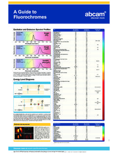
A Guide to Fluorochromes - Abcam
docs.abcam.comEnergy is then released in the form of a photon (fluorescence) and the electron moves back down to the lower energy level (Gs) (3). In the case of a tandem fluorescent dye, after excitation of the electron by a laser (1 - 2), energy is released …

Used Cooking Oil (UCO) as biofuel feedstock in the EU
www.transportenvironment.org1 Introduction 8 1.1 Background 8 1.2 Objective 8 1.3 Methodology and scope 8 1.4 Content of this report 9 ... LIF Laser-induced fluorescence NMR Nuclear Magnetic Resonance RED Renewable Energy Directive PYGCMS Pyrolysis Gas Chromatography Mass spectrometer UCO Used Cooking Oil ...

Leica TCS SP8
www.leica-microsystems.comof fluorescence spectra ... of older detection principles, such as pixel convolution, noise, and voltage-related scaling issues. backed by lightning-fast counting electronics, even ... microscopy has an inherent limitation of serial image collection. Camera-based
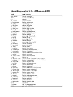
Quest Diagnostics Units of Measure (UOM)
www.questdiagnostics.commean intensity fluorescence units . Million/mL : million per microliter . min : minute . mL : milliliter . mL/24 h : milliliter per 24 hours . mL/min : per minute . milliliter mL/min/1.73m2 : nute per 1.73 meters squared . milliliter per mi mm : millimeter . mm/h : millimeter per hour . mm/Hg : millimeters of mercury . mm2/mm3 : per cubic ...

DAPI Nucleic Acid Stain - Thermo Fisher Scientific
assets.thermofisher.comSample Preparation Use the fixation protocol appropriate for your sample, or use the following protocol. 2.1 5 6Collect a cell suspension of 2 × 10 to 1 × 10 cells. 2.2 Pellet the cells by centrifugation and discard the supernatant. Figure 1. Fluorescence excitation and emission profiles of DAPI bound to dsDNA.
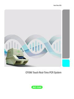
CFX96 Touch Real-Time PCR System - Bio-Rad Laboratories
www.bio-rad.comcombination designated for single-color fluorescence resonance energy transfer (FRET) experiments, further expanding your experimental options. Discrete excitation and detection wavelengths for the CFX96 Touch System enable thorough data discrimination. 425 450500 550 600 650 700 750 775 Detection Channel 1 510–530 FAM Channel 2 560–580 HEX

Chapter 3 Flame Atomic Absorption and Emission …
www.whitman.edulight is reflected by mirrors into the monochromator. This reflected light that contains various wavelengths past through a small slit that is size adjustable. ... through resonance fluorescence that emits a specific wavelength of UV or visible radiation (Section 2.2.2). This emission usually corresponds to only one or a few

IPC-4552A: Performance Specification for Electroless ...
www.ipc.orgX-Ray Fluorescence (XRF) Spectrometry [IPC-TM-650, Method 2.3.44]..... 90 APPENDIX 11 Phosphorus Content Measure-ment in ENIG Using Electron Dispersive Spectroscopy EDS – Initial Testing..... 97 APPENDIX 12 Standard Developments …

BD FACSCanto II
www.bd.comwhich data is less resolved but is acquired more quickly. A lower flow rate is generally used in applications for which optimal resolution and sensitivity are critical. Flow cell 5 ... direct light scatter and fluorescence signals through spectral filters to the detectors. Excitation The excitation optics consist of multiple fixed wavelength

06 DNA sequencing - California State University, …
www.csus.eduThe gel is dried onto chromatography paper and exposed to X-ray film. Since the template strand is not radioactively labeled, it does not generate a band on the X-ray film. ... the 4 different kinds of products are detected and the fluorescence intensity translated into a data “peak”. Thus all four chain termination reactions can be ...

User Guide: Qubit dsDNA HS Assay Kits
assets.thermofisher.comIntroduction: The Qubit ... RNA (Figure 1, page 7) and is accurate for initial sample concentrations from 10 pg/µL to 100 ng/µL. The assay is performed at room temperature, and the signal is stable for 3 hours. Common contaminants such as salts, free nucleotides, solvents, ... fluorescence; the Qubit ...

Relative quantification
www.gene-quantification.deconstant level of fluorescence or C P acquisition according to the established mathematic algorithm (see Section 3.6). Three general procedures of calculation of the relative quantification ratio are established: 1. The so-called ‘delta C t’ (eqs. 1–2 using ∆C P) or ‘delta-delta C t’ method (eqs. 3–4 using ∆∆C

EdU (5-ethynyl-2’-deoxyuridine)
tools.thermofisher.comFluorescence microscopy (Imaging) 50 coverslips Alexa Fluor® 488 dye C10337 • Includes blue-fluorescent nuclear counterstain, Hoechst 33342 • Not interchangeable with flow cytometry assays Alexa Fluor® 555 dye C10338 Alexa Fluor® 594 dye C10339 Alexa Fluor® 647 dye C10340 Table 4. Dye- and hapten-containing azides.

Fluorescence Excitation and Emission Fundamentals
www.chem.uci.eduFluorescence is the property of some atoms and molecules to absorb light at a particular wavelength and to subsequently emit light of longer wavelength after a brief interval, termed the fluorescence lifetime. The process of phosphorescence occurs in a manner similar to fluorescence, but with a much longer excited state lifetime.
Similar queries
Fluorescence, Fluorescence lifetime measurements, Resonance energy transfer, De la spectroscopie de fluorescence : de la, Quantique, Principles of Fluorescence and Fluorescence Microscopy, Breakthrough, Bio-Rad Laboratories, Virology Techniques, Microscopy, Basic Principles, Procedure Manual, Introduction, AFB Smear Microscopy, Fluorescence microscopy, Determination of Relative Fluorescence Quantum, Fluorescence spectroscopy, Sample Preparation, Photoluminescence, Energy, Fluorescence resonance energy, Dots for two-photon fluorescence imaging, Multimode, Fl uorescence, Energy transfer, Light, Fluorometer, Spectroscopy, Western Kentucky University, FlowJo Basic Tutorial, Of fluorescence, Energy Dispersive X-ray Spectroscopy EDS, Resolved fluorescence, X ray Fluorescence, Resonance, Principles, Quest Diagnostics, Sample, CFX96 Touch Real-Time PCR System, Fluorescence resonance, Atomic Absorption and Emission, Reflected, Reflected light, Resolved, DNA sequencing, California State University,, Procedures