Transcription of AFB Smear Microscopy - APHL
1 AFB Smear Microscopy 1. Terminology AFB Smear Microscopy : Microscopic examination of specially stained smears to detect acid-fast organisms such as Mycobacterium tuberculosis and non- tuberculous mycobacteria (NTM). Acid Fast Bacilli (AFB): organisms (including mycobacteria) that resist decolorization with acid alcohol due to the lipid-rich mycolic acids in the cell wall thereby retaining the primary stain 2. Terminology Processing: digestion, decontamination, and/or concentration of a primary patient specimen prior to setting up culture and Smear Smear : A small amount of primary patient specimen (direct or processed) is placed on a slide for the purpose of microscopic examination 3.
2 AFB Microscopy Examination of smears is a rapid, convenient and inexpensive test All types of specimens can be evaluated sputum, tissue, body fluids, etc. Positive AFB Smear results provide a first indication of mycobacterial infection and potential TB disease Must be accompanied by additional testing including culture for confirmatory diagnosis 4. AFB Microscopy Results Guide Decisions Clinical management Patient therapy may be initiated for TB based on Smear result and clinical presentation Changes in Smear status important for monitoring response to therapy Laboratory testing Algorithms for use of nucleic acid amplification tests are often based on Smear positivity Public health interventions Smear status and grade useful for identifying the most infectious cases Contact investigations prioritized based on Smear result Decisions regarding respiratory isolation based on Smear result 5.
3 Smear -positive TB Cases Smear -positivity and grade indicates relative bacterial burden and correlates with disease presentation Patients that are sputum Smear -positive are 5 10. times more infectious than Smear negative patients Untreated or treated with an inappropriate regimen, a sputum Smear -positive patient may infect 10-15. persons/year 6. Sputum Smear Results In 2010, 43% of pulmonary TB cases in the were sputum Smear positive Incremental diagnostic yield of examination of three sputum specimens among Smear positive cases First specimen Second specimen Third specimen CDC. Reported Tuberculosis in the United States, 2010. Atlanta, GA: Department of Health and Human Services, CDC, October 2011.
4 7 in Mase S, Ramsay A, Ng N, Henry M, Hopewell PC, Cunningham J, Urbanczik R, Perkins M, Aziz MA, Pai M. Yield of serial sputum specimen examinations the diagnosis of pulmonary tuberculosis: a systematic review. Int J Tuberc Lung Dis 2007;11(5):485-95. Limitations of AFB Microscopy Does not distinguish between viable and dead organisms Follow-up specimens from patients on treatment may be Smear positive yet culture negative Limited sensitivity High bacterial load 5,000-10,000 AFB /mL is required for detection Misses >45% of TB cases Limited specificity All mycobacteria are acid fast Does not provide species identification Local prevalence of MTB and NTM determine the 8.
5 Predictive values of a positive Smear for MTB. Smear Types Direct Smear Smear prepared directly from a patient specimen prior to processing Concentrated Smear Smear prepared from a processed specimen after centrifugation is used to concentrate the material 9. Direct Smears Rapid and simple May be performed for quick results Positive results help confirm clinical suspicions Not as sensitive as concentrated Smear A direct Smear should always be followed by a concentrated Smear 10. Concentrated Smear 3-10 ml of sputum is processed and concentrated by centrifugation Smears can be made Directly from processed pellet May increase Smear sensitivity However, may result in less material available for NAA testing & culture From re-suspended pellet after the addition of buffer Re-suspended in ~2 ml buffer; one drop for Smear 11.
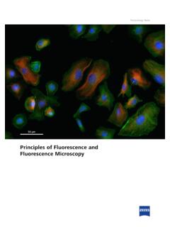
6 AFB Microscopy Staining Techniques Two basic techniques: same principle: fluorescence : Auramine staining Also known as Fluorochrome staining Contrast light & dark Brightfield: carbol fuchsin staining Contrast red AFB on blue or green background 12. Carbol-Fuchsin AFB Staining Primary stain is Carbol Fuchsin Ziehl-Neelson (ZN). Requires heat during staining Requires higher magnification More fields examined ( 300 fields at 1000X). Requires more time to read Requires use of oil immersion Stains all NTM well Kinyoun Cold carbol fuchsin method Less toxic fumes Neither method is reccomended for staining primary specimens 13. Fluorescent AFB Staining Primary stain is Fluorescent CDC recommends fluorochrome staining for detecting AFB in primary patient specimens Auramine-O, Auramine Rhodamine Read at lower magnification, less fields examined ( , 30 fields at 200X).
7 Faster screening of smears than with ZN. ~10% more sensitive than ZN. Does not require use of oil immersion 14. Steps in Performing AFB. Microscopy 1. Preparing and Fixing Smears 2. Staining Smears 3. Examining Smears 4. Recording and Reporting Results 15. Getting Started . New, clean, greaseless, and unscratched slides should be used Match identifiers on slide with clinical specimens Labeling should be performed with material that stays permanently affixed to the slide during the staining procedure ( , graphite or diamond tip pencil). 16. 17. Preparing Smears After processing and concentrating the specimen, 1 to 2 drops of material should be smeared on the slide Smear material in an area of approximately 2-cm2.
8 18. Too Just Too Thick Right Thin 19. Fixing Smears Prior to heat fixing, smears should be allowed to air dry completely within the biological safety cabinet Acceptable methods for heat fixing Flame fixing by passing over flame 2 3 times for a few seconds Smear side up Avoid scorching 2 hrs minimum at 65-75oC on an electric slide warmer 15 min at 80oC1. 5% phenol in 70% EtOH for 5 min2. also kills AFB. Considerations Flame fixing may aerosolize organisms from Smear Insufficient heat or time can lead to Smear washing off Slide warmers may not heat evenly Viable AFB remain with some fixing methods 1 Bailey & Scott's Diagnostic Microbiology 2007 12th ed.
9 2 Chedore et al. 2002. J. Clin. Microbiol. 40:4077 20. CLSI M48-A: Laboratory Detection & Identification of Mycobacteria Keep slides on warmer for 2 hours 21. Heat Fixing Smear Safety Concerns: Wear gloves at all times Work inside the BSC. Use open flame for shor periods of time 22. AFB Staining Principles Primary stain penetrates cell wall Intense decolorization does not release primary stain from the cell wall of AFB. Color of AFB-based on primary stain Counterstain provides contrasting background 23. Stains Used in fluorescence Microscopy Primary Stains Auramine O. Auramine O-Rhodamine-B. Counter Stains Potassium Permanganate Acridine Orange 24.
10 fluorescence AFB Microscopy Primary fluorochrome Auramine O. Auramine O-Rhodamine B. Counter Stain Potassium permanganate 25. fluorescence AFB Microscopy Primary Fluorochrome AFB Fluoresces Auramine O Green Auramine O/ Yellow/Orange Rhodamine B. Acridine Orange Yellow/Orange 26. 450x 27. Water Quality is Key AFB Microscopy is not specific Acid fast environmental contaminants as well as NTM and MTB presence in the specimen will be detected Introduction of an environmental contaminant during wash steps and in reagent preparation must be avoided Use of a negative control slide essential for detecting potential environmental contaminants Avoid using large containers of reagents and carboys of water Use filtered distilled or deoinized water Water filtration and distribution systems should be monitored 28.