Flow Doppler
Found 10 free book(s)
US Carotid Doppler - UT Southwestern Medical Center
www.utsouthwestern.eduo Color-flow Doppler images with proper color scale to demonstrate areas of high flow and color aliasing o Spectral Doppler gains will be set to allow a spectral window and optimized to reduce artifact o An angle of 60 degrees or less will be used to measure velocities o Doppler angle should always be parallel to the vessel wall

Billing and Coding Guidelines AMA CPT/ ADA CDT Copyright ...
downloads.cms.govand with color flow doppler echocardiography C8930 Transthoracic echocardiography with contrast, or without contrast followed by with contrast, real-time with image documentation (2D), includes M-mode . recording, when performed during rest and cardiovascular stress test using

4.2 Instrumentation: Pressure, Flow, & Level - Fermilab
uspas.fnal.gov4. Acoustic flow meters based on Doppler effect 5. Turbine flow meters where frequency ~ velocity 6. Optical techniques (Laser Doppler) Single phase flows Two phase flows 1. Void fraction measurement (A v/A) a) Capacitance measurement b) Optical characterization 2. Quality measurement (m v/m) These techniques are for

Ultrasound Examination of the Extracranial Cerebrovascular ...
www.aium.orgColor Doppler is also helpful to detect external carotid artery branches to definitively identify this vessel. Color Doppler should be used to clarify the cause of image/pulsed Doppler mismatches and to detect narrow flow channels at sites of stenosis.9 Power Doppler evaluation may be complementary to color Doppler to search for a

Echo in Pulmonary HTN - asecho.org
asecho.orgOct 10, 2017 · •Diastolic flow predominance in hepatic veins. PW Doppler from Hepatic Vein SFF = systolic filling fraction (%) = S ... Echo Average of Echo-Doppler and RHC Measurement-and Measurement Excellent and good quality Doppler signal Fair and poor quality Doppler signal Mathai Advances in Pulm Hypertension 2008;7:1-7.

General principles of carotid Doppler ultrasonography
www.e-ultrasonography.orgFig. 1. Typical Doppler spectrum of the internal carotid artery and the external carotid artery. A. The Doppler spectrum of the internal carotid artery shows a low resistance pattern with sufficient diastolic antegrade flow. B. The external carotid artery shows a more resistive pattern than the internal carotid artery.

Surgical and Ablative Procedures for ... - UHCprovider.com
www.uhcprovider.comanatomical structure while Doppler detects the flow, direction of flow and flow velocity. Endovenous Ablation : A minimally invasvi e procedure that uses heat generated by radiofrequency (RF) or laser energy to seal off damaged veins.
Doppler Ultrasound - Principles and practice - Fetal Medicine
fetalmedicine.orghigher-frequency Doppler signals. The beam/flow angle at (C) is almost 90° and there is a very poor Doppler signal. The flow at (D) is away from the beam and there is a negative signal. All types of Doppler ultrasound equipment employ filters to cut out the high amplitude, low-
Doppler ultrasonography of the lower extremity arteries ...
www.e-ultrasonography.orgDoppler US is a good method for screening and follow-up, as well as for the definitive diagnosis of peripheral arterial disease [3-7]. Color Doppler US can easily identify arteries by finding round objects with regular pulsation and can be used to detect stenotic or occluded segments [4,8]. Pulsed-wave Doppler US can show the exact flow velocity of
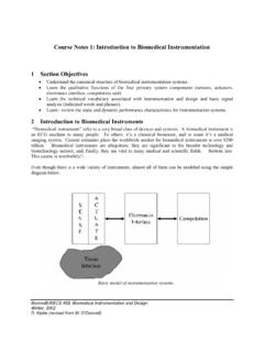
Course Notes 1: Introduction to Biomedical Instrumentation ...
www.eecs.umich.eduflow dynamics in an artery (e.g., ultrasound color flow imaging of the carotid artery) is an example of a non-invasive sensor. Contact/Remote: A strain gauge sensor attached to a muscle fiber can record deformations and forces in the muscle. An MRI or ultrasound imaging system can measure internal deformations and forces without