Search results with tag "Compound microscope"

BIOLOGY Grade 9 - Science
msschultzteaching.weebly.comIntroduction . Early History of the Microscope The Parts of the Compound Microscope . Taking Care of the Microscope Requirements for Obtaining a Good Image . Finding an Object under the ... Microscope: Compound Microscopes of Today How to Use the Compound Microscope: Objective Len s Choice .

AN INTRODUCTION TO THE COMPOUND MICROSCOPE
users.rowan.eduAN INTRODUCTION TO THE COMPOUND MICROSCOPE OBJECTIVE: In this lab you will learn the basic skills needed to stain and mount wet slides. You will also learn about magnification, resolution and the parts of the compound microscope. INTRODUCTION: The light microscope can extend our ability to see detail by 1000 times, so that we can
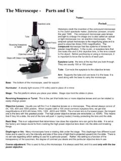
The Microscope Parts and Use - Plainview
www.pobschools.orgthe year 1590. The compound microscope uses lenses and light to enlarge the image and is also called an optical or light microscope (vs./ an electron microscope) . The simplest optical microscope is the magnifying glass and is good to about ten times (10X) magnification. The

LAB 4 Microscopy & Cells
www.lamission.eduExplain each part of the compound microscope and its proper use. 2. Examine a variety of cells with the compound microscope and estimate cell size. 3. Examine larger specimens with the stereoscopic dissecting microscope. Introduction In this laboratory you will be learning how to use one of the most important tools in biology – the
Cell Biology, Molecular Biology And Biotechnology
www.uou.ac.inThe first useful compound microscope was invented in 1590 by Francis Janssen and Zacharias Janssen. Their microscope had two lenses and total magnifying power between 10X and 30X. Such types of microscopes were called ―flea glasses", since they were primarily used to examine small whole organisms such as fleas and other insects.

Lab 4 - Comparison of Parasitic and Free-Living Worms
www.amherst.edu3 B) Dugesia, microscope slide (Figure 2). Observe a prepared whole mount of Dugesia under low power of your compound microscope. You should see the eye spots and the diverticula of the gut. Also look for the "brain", nerve cord, and excretory system. C) Clonorchis, Class Trematoda, preserved specimen and microscope slide (Figure 3). Clonorchis sinensis, the …
Microscope Instruction Manual - Fisher Sci
static.fishersci.comThe compound microscope combines two optical lens systems. The lens closest to the specimen slide, the objective, magnifies the primary image and the top lens, called the eyepiece, further magnifies the image. Magnification of the objective …