Transcription of 10. Pharyngeal Arches Revisited and the …
1 10-1 Letty Moss-Salentijn DDS, PhDDr. Edwin Professor of Dentistry (in Anatomy and Cell Biology)E-mail: ASSIGNMENT: Larsen 3rd edition: ; pp. 371-378; pp. 393 bottom-398 SUMMARY:The transient structures that are known as Pharyngeal grooves and Pharyngeal pouches disappear toward theend of the embryonic period. The first Pharyngeal groove will give rise to the external auditory meatus of theadult ear. The other three grooves will disappear without having any further role in the development of Pharyngeal pouches develop into a series of structures that include the pharyngotympanic tube, middle earcavity, palatine tonsil, thymus, the four parathyroid glands, and the ultimobranchial bodies of the thyroid related development of the external ear by contributions of the first and second Pharyngeal Arches isreviewed, as is the development of the tongue, which is formed by contributions from the first throughfourth Arches .
2 Finally, the development of the thyroid gland, which is spatially related to the derivativesof the third and fourth Pharyngeal pouches, is discussed OBJECTIVES You should be able to:a. List the derivatives of the first Pharyngeal groove and describe the pattern of obliteration ofpharyngeal grooves List the derivatives of the different Pharyngeal Describe briefly the development of the external and middle List the different swellings and explain the process of merging of these swellings in thedevelopment of external ear and tongue. You should be able to list the Pharyngeal Arches thatcontribute to these Describe the sequence of events that lead to the development of the palatine Describe the initial development of the thyroid gland, list the initial site of development anddescribe the pathway of the growing gland.
3 Describe the thyroglossal Describe the origin and developmental movements of the thymus gland and explain how theparathyroid glands, which develop from the third pouch become inferior while those thatdevelop from the fourth pouch become superior . Arches Revisited and thePHARYNGEAL POUCHES10-2 GLOSSARY:Auricle. External ear, derived from three swellings (hillocks) on the first arch and three swellings on theapposing surface of the second A median swelling on the Pharyngeal surface of the second Pharyngeal Arches . Is obliterated duringfurther tongue operculum. A large outgrowth of the second Pharyngeal arch, which obliterates the second, third andfourth Pharyngeal eminence. A combined median swelling on the Pharyngeal surface of the combined third andfourth Pharyngeal Arches .
4 This swelling will give rise to the posterior third of the tongue and the duct. The duct that initially runs between the developing thyroid gland and the surface of thetongue, where it opens at the foramen cecum. The duct soon loses its lumen and becomes a solid cord: thethyroglossal tract. See: Thyroglossal crypts. Epithelially lined depressions that extend into the underlying tonsillar fossa. A small recess of the ventral second Pharyngeal stroma. Subepithelial connective tissue, which is infiltrated by lymphoid impar. Median swelling on the Pharyngeal surface of the first Pharyngeal Arches . Participates in theformation of the anterior 2/3 of the :As was seen before, the Pharyngeal Arches are bilateral/paired swellings that surround the foregut of theembryo and develop in a rostral to caudal sequence, in the fourth and fifth week of development.
5 They arewedged between the developing heart and brain. On the ectodermal (future skin) surface, the depressionsbetween the Pharyngeal Arches are called Pharyngeal the endodermal (future Pharyngeal )surface the depressions between the Pharyngeal Arches are called Pharyngeal ectodermally lined Pharyngeal grooves are transient in nature and of limited the first Pharyngeal groove gives rise to a permanent structure: the ear canal or external auditorymeatus. The developmental sequence will be described below. The second, third and fourth grooves arerapidly covered by a large outgrowth of the second Pharyngeal arch, the hyoid operculum, andobliterated ( ).In contrast, the endodermally lined Pharyngeal pouches, while also transient in nature, all giverise to important structures.
6 In reviewing the derivatives of the Pharyngeal pouches you must keep inmind that each pouch has a dorsal and a ventral part ( ):pouch 1ventralobliterated: no derivativedorsalpharyngotympanic tube and middle ear cavitypouch 2ventralpalatine tonsil, partially obliterated by endodermdorsalcontributes to pharyngotympanic tubepouch 3ventralthymusdorsalinferior parathyroid glandpouch 4ventral ultimobranchial bodies (C-cells of thyroid gland)dorsalsuperior parathyroid gland By the middle of the eighth week all Pharyngeal pouch derivatives have reached the Fate of the Pharyngeal grooves. The first Pharyngeal groove forms the external auditory meatus. The secondpharyngeal arch expands and fuses with the cardiac eminence to cover the remaining Pharyngeal grooves, which form thetransient lateral cervical ROLE OF FIRST Pharyngeal POUCH AND GROOVE IN EAR DEVELOPMENT(Fig.)
7 10-3)We distinguish external, middle and internal ear. The development of the internal ear from theotic placode will be discussed in lecture development of the external ear involves contributions of Pharyngeal Arches 1 and 2 to form theauricle, and the first Pharyngeal groove to form the external auditory meatus. The auricle is derived from threeswellings (hillocks) on the first arch and three swellings on the opposing surface of the second. Thedeveloping external auditory meatus lies in between. The swellings gradually are molded together (bymerging) to form the characteristic shape of the auricle ( ). The external ear can be a usefuldiagnostic tool: if it is severely malformed, you should anticipate problems in the other derivatives ofthe first and second development of the middle ear involves skeletal elements of Pharyngeal Arches 1 and 2 that formthe three ossicles of the middle ear: incus, malleus and stapes, and the first Pharyngeal pouch to formthe lining of the middle ear cavity.
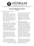
8 The auditory ossicles begin forming during the 7th week. The dorsal10-4 Fig. 10-3. Development of the ear. The components of the inner, middle, and outer ears arise in coordination from severalembryonic structures. The otic vesicle gives rise to the membranous labyrinth of the inner ear and to the eighth nerve , B, The superior end of the otic vesicle forms an endolymphatic appendage, and the body of the vesicle then differentiatesinto utricular and saccular regions. C, D, The endolymphatic appendage elongates to form the endo-lymphatic sac and duct;the utricle gives rise to the three semicircular ducts; and the inferior end of the saccule elongates and coils to form thecochlear duct. Simultaneously, the three auditory ossicles arise from mesenchymal condensations formed by the first andsecond Pharyngeal Arches ; the first Pharyngeal pouch enlarges to form the tubotympanic recess (the future middle ear cavity),and the first Pharyngeal cleft (the future external auditory meatus) becomes filled with a transient meatal plug of ectodermalcells.
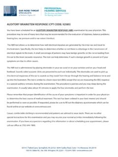
9 C, Finally, in the ninth month, the tubotympanic cavity expands to enclose the auditory ossicles, forming the functionalmiddle ear cavity. The definitive eardrum represents the first Pharyngeal membrane and is thus a three-layered structurecomprising ectoderm, mesoderm, and Pharyngeal pouch elongates further dorsally to form a tubotympanic recess, which becomes thepharyngotympanic tube and the middle ear cavity (tympanic cavity). In this cavity the firstpharyngeal pouch epithelium extends to surround the ossicles. Where the first Pharyngeal pouchepithelium meets the epithelium of the first Pharyngeal groove, the tympanic membrane or 10-4. Differentiation of the auricle. The auricledevelops from six aricular hillocks, which arise onthe apposed surfaces of the first and secondpharyngeal Arches .
10 (A, Photo courtesy of Dr. ArnoldTamarin.)SECOND Pharyngeal POUCHThe tonsil forms in a small persisting recess - the tonsillar fossa - of the ventral pharyngealpouch, which is almost completely obliterated by the proliferation of the endodermal lining. Thepalatine tonsil - the tonsil - develops during the third month as a subepithelial infiltration of lymphoidtissue: tonsillar stroma. Solid cellular strands from the overlying epithelium subsequently extend intothis stroma. The endothelium and associated mesenchymal stroma constitute the primordium of thetonsil. The cellular strands gradually open up to form tonsillar crypts. At about the fifth monthlymphatic aggregates, coming from the blood stream, appear in the tonsillar stroma.