Transcription of Acceptance Testing of a Digital Breast Tomosynthesis Unit
1 3/29/12. Acceptance Testing of a Digital Breast Tomosynthesis unit 2012 AAPM Spring Clinical Meeting Jessica Clements, , DABR. Objectives Review of technology and clinical advantages Acceptance Testing Procedures QC Testing Procedures Accreditation Process Selenia Dimensions 2D Digital Mammography System 3D Digital Breast Tomosynthesis Received FDA Approval in February of 2011. For existing 2D system, a software upgrade is required to activate DBT. DBT is considered a new mammographic modality separate from Full Field Digital Mammography Image from the Dimensions QC Manual 1. 3/29/12. unit design Allows both 3D and 2D images to be acquired under the same Breast compression Improved selenium detector, 70 m pixel size High Transmission Cellular (HTC) anti-scatter grid in 2D.
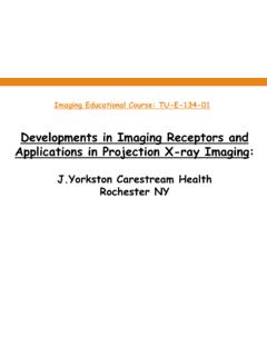
2 Tungsten target x-ray tube Filter options Rh and Ag (2D). Al (3D). 2D Breast Imaging Single projection results in tissue superimposition that is created by the overlap of normal Breast structures Slide courtesy of Hologic 3D Principle of Operation Arc of motion of x-ray tube, showing individual exposures X-ray tube moves in an arc across the Breast A series of low dose images are acquired from different angles Total dose approximately the same as one 2D. mammogram Projection images Compression Paddle are reconstructed Compressed into 1 mm slices Breast {. Reconstructed Slices Detector Housing Slide courtesy of Hologic 2. 3/29/12. 1. The x-ray tube contains a tungsten target and the following material is the filter used in 3D imaging: 6% 1.}
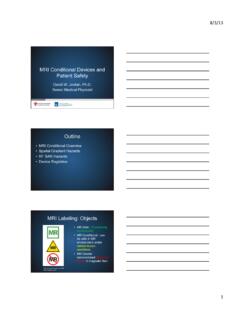
3 Tungsten 13% 2. silver 81% 3. aluminum 0% 4. rhodium 0% 5. molybdenum Correct Answer 1. tungsten not in unit 2. silver 2D only 3. aluminum 4. rhodium 2D only 5. Molybdenum not in unit Reference: Ren, B., Ruth, C., Wu, T., Zhang, Y., Smith, A., Niklason, L, A new generation FFDM/ Tomosynthesis fusion system with selenium detector , Proc. SPIE 7622, 76220B (2010). *video of combo exam of ACR Phantom 3. 3/29/12. Images courtesy of Hologic 2D Mammogram Tomosynthesis Better Sensitivity Slide courtesy of Hologic 2D Mammogram Tomosynthesis Fewer Recalls Slide courtesy of Hologic 4. 3/29/12. Advantages Imaging dense Breast tissue May decrease recall rate Addresses issue of tissue overlap Dose within existing limits for 2D.
4 Mammography Personnel Requirements Required Training for a medical physicist: Digital Mammography: MQSA Personnel requirements including 8 hours of training in surveying units of Digital mammography Digital Breast Tomosynthesis : Must receive at least 8 hours of training in Digital Breast Tomosynthesis modality Responsibilities unit must be evaluated by a medical physicist before clinical use commences after the equipment is first installed, moved, or significantly modified The medical physicist must review the results of the technologist's QC tests at least annually. The results of the overall Quality Assurance and Quality Control program must be reviewed by the medical physicist with the responsible interpreting physician at least annually.
5 5. 3/29/12. Acceptance Testing Required in the following situations: New unit Upgrade of 2D to include DBT. After major component upgrades including software, detector, tube, etc. Release notes will indicate what tests must be performed after manufacturer upgrades Quality Control Manual Quality Control Tests to be Performed by the Medical Physicist Upon Installation Table 1-1 QC Manual *Mammographic unit Assembly Collimation Assessment Evaluation Artifact Evaluation *kVp Accuracy and Reproducibility *Beam Quality Assessment HVL Evaluation of System Resolution AEC Function Performance Breast Entrance Exposure, AEC. Reproducibility and AGD. Radiation Output Rate Phantom Image Quality Evaluation Signal to Noise and Contrast to Noise Diagnostic Review Workstation Quality Control DICOM Printer Quality Control Detector Flat Field Calibration Geometry Calibration for Compression Thickness Indicator Tomosynthesis Option *Compression *Described in the 1999 ACR Mammography QC Manual 6.
6 3/29/12. Action Categories Technologist QC to be performed by the medical physicist at Acceptance 7. 3/29/12. Detector Flat Field Calibration Pre-programmed set of exposures of the flat-field acrylic phantom without a compression paddle Review preview images for foreign objects, gross artifacts, or other non- uniformities or collimation interference Performed weekly by technologist Category A. Geometry Calibration for Tomosynthesis Image the provided geometry phantom as specified Evaluated automatically by software in the system Performed by technologist semi- annually Category A. Geometry Calibration for Tomosynthesis 8. 3/29/12. Compression Thickness Indicator Compress the cm spot contact compression paddle onto the ACR.
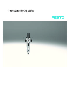
7 Phantom using full automatic compression of approximately 30. pounds Record the thickness indicated Performed by the technologist bi-weekly Category C. DICOM Printer QC. A SMPTE pattern is printed from the imaging system and optical densities are measured. Mid density (40%), lower density (90%), and density difference (10%-40%). Limit: + Performed by the technologist weekly Category B. DICOM Printer QC. 9. 3/29/12. Medical Physicist QC Tests Collimation Measure the deviation between x-ray field and edges of the image receptor: 24x29, 18x24 (L), 18x24 (C), 18x24 (R), and 18x29 cm tomo Measure distance with Digital ruler X-ray field must not extend by more than 2% SID on any side of the detector Category C. Image from the Dimensions QC Manual Collimation Measure the deviation between x-ray field and light field: 24x29 cm paddle Use ready pack, GafChromic film, or other test tool to visualize x-ray field The total misalignment of the edges of the light field and the x-ray field along either the length or the width of the visually defined field at the plane of the Breast support surface must not exceed 2% of the SID.
8 Category C. 10. 3/29/12. Collimation Measure the deviation between compression paddle and edges of the image receptor: 24x29, 18x24 cm, and small Breast (if available). The anterior edge of the compression paddle must be aligned just beyond the chest wall edge of the image receptor so that it does not appear in the mammogram. The anterior edge must not extend beyond the chest wall edge of the image receptor by more than 1% of the SID. Category C. Beam Quality Assessment Follow the ACR manual Protect the detector with lead To measure in 3D mode, use zero degree tomo mode Only minimum HVL limits apply Category C. Artifact Evaluation Image the flat field phantom (4 cm thick acrylic block manufacturer provided).
9 No compression paddle All filters in each imaging mode (conventional, tomo, magnification). Rotate phantom 180* between exposures View full resolution images for artifacts Print flat field pattern as 2560x3328 for 8x10 . film and 3328x4096 for 24x30 cm film, both as True Size Printing Category C. 11. 3/29/12. Image courtesy of Hologic Artifact Examples Dead pixels cluster of 16 or more complete or partial row or column System Resolution Uses system limiting spatial resolution as performance indicator 18x24 cm compression paddle High contrast resolution pattern that can test up to 15 lp/mm with 1 lp/mm steps from 3-15 lp/mm Pattern is placed on flat field phantom approximately 1 cm from chest wall edge, with pattern angled 45* to the anode- cathode axis 12.
10 3/29/12. Image courtesy of Hologic Image courtesy of Hologic System Resolution In 2D, limiting resolution must be greater than 7 lp/mm In 3D, must be greater than 3 lp/mm Category A. 13. 3/29/12. 3. When the test pattern is approximately 45 to the anode- cathode axis and when imaging in Tomosynthesis mode, the system limiting spatial resolution must be greater than: 82% 1. 3 lp/mm 0% 2. 5 lp/mm 18% 3. 7 lp/mm 0% 4. 9 lp/mm 0% 5. 11 lp/mm Correct Answer 1. 3 lp/mm 2. 5 lp/mm 3. 7 lp/mm minimum in 2D mode 4. 9 lp/mm 5. 11 lp/mm Reference: Hologic: Quality Control Manual Selenia Dimensions 2D FFDM Selenia Dimensions DBT, 2011 (Part Number MAN-01965 Revision 002) p. 35. AEC Function Performance Record exposure index (and exposure parameters) for phantom thicknesses (BR-12 or acrylic) in various AEC modes used clinically in conventional, tomo, and magnification modes.