Transcription of ACR Thyroid Imaging, Reporting and Data System (TI-RADS ...
1 ORIGINAL ARTICLE HEALTH SERVICES RESEARCH AND POLICY. ACR Thyroid imaging , Reporting and data System (TI-RADS): White Paper of the ACR TI-RADS Committee Franklin N. Tessler, MD, CM a , William D. Middleton, MD b, Edward G. Grant, MD c, Jenny K. Hoang, MBBS d, Lincoln L. Berland, MD a, Sharlene A. Teefey, MD b, John J. Cronan, MD e, Michael D. Beland, MD e, Terry S. Desser, MD f, Mary C. Frates, MD g, Lynwood W. Hammers, DO h,i, Ulrike M. Hamper, MD j, Jill E. Langer, MD k, Carl C. Reading, MD l, Leslie M. Scoutt, MD m, A. Thomas Stavros, MD n Abstract Thyroid nodules are a frequent nding on neck sonography. Most nodules are benign; therefore, many nodules are biopsied to identify the small number that are malignant or require surgery for a de nitive diagnosis. Since 2009, many professional societies and in- vestigators have proposed ultrasound-based risk strati cation systems to identify nodules that warrant biopsy or sonographic follow-up.
2 Because some of these systems were founded on the BI-RADS classi cation that is widely used in breast imaging , their authors chose to apply the acronym TI-RADS, for Thyroid imaging , Reporting and data System . In 2012, the ACR convened committees to (1) provide recommendations for Reporting incidental Thyroid nodules, (2) develop a set of standard terms (lexicon) for ultrasound Reporting , and (3). propose a TI-RADS on the basis of the lexicon. The committees published the results of the rst two efforts in 2015. In this article, the authors present the ACR TI-RADS Committee's recommendations, which provide guidance regarding management of Thyroid nodules on the basis of their ultrasound appearance. The authors also describe the committee's future directions. Key Words: Thyroid nodule, Thyroid cancer, management guidelines, ultrasound J Am Coll Radiol 2017;-:---. Copyright 2017 American College of Radiology INTRODUCTION is the most effective, practical test to determine whether a Thyroid nodules are exceedingly common, with a reported nodule is malignant or may require surgery to reach a prevalence of up to 68% in adults on high-resolution de nitive diagnosis [2].
3 However, most nodules are ultrasound [1]. Currently, ne-needle aspiration (FNA) benign, and even malignant nodules, particularly ones a j Department of Radiology, University of Alabama at Birmingham, Department of Radiology and Radiological Science, Johns Hopkins Birmingham, Alabama. University, School of Medicine, Baltimore, Maryland. b k Mallinckrodt Institute of Radiology, Washington University School of Department of Radiology, University of Pennsylvania, Philadelphia, Medicine, St Louis, Missouri. Pennsylvania. c l Department of Radiology, Keck School of Medicine, University of Department of Radiology, Mayo Clinic College of Medicine, Rochester, Southern California, Los Angeles, California. Minnesota. d m Department of Radiology, Duke University School of Medicine, Durham, Department of Radiology and Biomedical imaging , Yale University, New North Carolina. Haven, Connecticut.
4 E n Department of Diagnostic imaging Brown University, Providence, Rhode Department of Radiology, University of Texas Health Sciences Center, Island. San Antonio, Texas. f Department of Radiology, Stanford University Medical Center, Stanford, Corresponding author and reprints: Franklin N. Tessler, MD, CM, California. Department of Radiology, University of Alabama at Birmingham, g Birmingham, AL 35249; e-mail: Department of Radiology, Brigham and Women's Hospital, Boston, Massachusetts. Dr Berland received personal fees from Nuance Communications during h the conduct of the study. Dr Beland has received personal fees from Hitachi Hammers Healthcare imaging , New Haven, Connecticut. i Department of Internal Medicine, Yale School of Medicine, New Haven, Aloka America outside the submitted work. All other authors have no Connecticut. con icts of interest related to the material discussed in this article.
5 2017 American College of Radiology 1546-1440/17/$ n 1. Downloaded for Anonymous User (n/a) at University of Saskatchewan - Canada Consortium from by Elsevier on April 13, 2017. For personal use only. No other uses without permission. Copyright 2017. Elsevier Inc. All rights reserved. smaller than 1 cm, frequently exhibit indolent or surveys, represent the consensus opinion of the ACR TI- nonaggressive behavior [3-5]. Therefore, not all detected RADS Committee. They are based on the literature;. nodules require FNA and/or surgery. analysis of data from the Surveillance, Epidemiology, and Despite a rapid increase in the reported incidence of End Results (SEER) Program of the National Cancer papillary Thyroid cancer that resulted from screening Institute; evaluation of existing risk classi cation systems;. Thyroid sonography in asymptomatic patients in South and expert opinion.
6 Our recommendations are intended to Korea, mortality has remained extremely low [6]. In the serve as guidance for practitioners who incorporate ultra- United States, overdiagnosis of Thyroid cancer, de ned sound in the management of adult patients with Thyroid as diagnosis of Thyroid tumors that would not, if left nodules. They should not be construed as standards. alone, result in symptoms or death accounted for 70% Interpreting and referring physicians are legally and pro- to 80% of Thyroid cancer cases in women and 45% of fessionally responsible for applying their professional cases in men between 2003 and 2007 [7]. judgment to every case, regardless of the ACR TI-RADS. Therefore, a reliable, noninvasive method to identify recommendations. The decision to perform FNA should which nodules warrant FNA on the basis of a reasonable also account for the referring physician's preference and the likelihood of biologically signi cant malignancy would be patient's risk factors for Thyroid cancer, anxiety, comor- highly desirable.
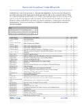
7 In 2015, committees convened by the bidities, life expectancy, and other relevant considerations. ACR published white papers that presented an approach to incidental Thyroid nodules and proposed standard termi- nology (lexicon) for ultrasound Reporting [8,9]. The OVERVIEW OF ACR TI-RADS. purpose of the present white paper is to present our The ultrasound features in the ACR TI-RADS are cate- System for risk strati cation, which is designed to identify gorized as benign, minimally suspicious, moderately most clinically signi cant malignancies while reducing the suspicious, or highly suspicious for malignancy. Points are number of biopsies performed on benign nodules. given for all the ultrasound features in a nodule, with more suspicious features being awarded additional points. PROJECT RATIONALE AND CONSENSUS Figure 1 presents these features arranged per the ve PROCESS lexicon categories [8].
8 When assessing a nodule, the Several professional societies and groups of investigators reader selects one feature from each of the rst four have proposed methods to guide ultrasound practitioners categories and all the features that apply from the nal in recommending FNA on the basis of ultrasound features category and sums the points. The point total [10-18]. Some of these systems were termed TI-RADS determines the nodule's ACR TI-RADS level, which ( Thyroid imaging , Reporting and data System ) because ranges from TR1 (benign) to TR5 (high suspicion of they were modeled on the ACR's BI-RADS , which has been malignancy). Note that although it is possible for a widely accepted in breast imaging . Other societies, such as the nodule to be awarded zero points and hence be charac- American Thyroid Association (ATA), have taken a slightly terized as TR1, all other nodules merit at least two points different, pattern-oriented approach, but with the same because a nodule that has a mixed cystic and solid intent [19].
9 The plethora, complexity, and lack of congruence composition (one point) will also gain at least one more of these systems has limited their adoption by the ultrasound point for the echogenicity of its solid component. Finally, community and inspired our effort to publish a classi cation although sonoelastography is a promising technique System under the auspices of the ACR. The ACR TI-RADS [20,21], it is probably not available in many ultrasound Committee agreed on the following attributes for our risk laboratories and is not incorporated into the ACR TI- classi cation algorithm. It would be: RADS. In the ACR TI-RADS, recommendations for FNA or n founded on the ultrasound features de ned in our ultrasound follow-up are based on a nodule's ACR TI- previously published lexicon;. RADS level and its maximum diameter. For risk levels n easy to apply across a wide gamut of ultrasound TR3 through TR5, the chart presents a size threshold at practices.
10 Or above which FNA should be recommended. We also n able to classify all Thyroid nodules; and de ned lower size limits for recommending follow-up n evidence based to the greatest extent possible. ultrasound for TR3, TR4, and TR5 nodules to limit The proposals presented in this white paper, which the number of repeat sonograms for those that are likely were developed via conference calls, e-mail, and online to be benign or not clinically signi cant. 2 Journal of the American College of Radiology Volume - n Number - n Month 2017. Downloaded for Anonymous User (n/a) at University of Saskatchewan - Canada Consortium from by Elsevier on April 13, 2017. For personal use only. No other uses without permission. Copyright 2017. Elsevier Inc. All rights reserved. ACR TI-RADS. COMPOSITION ECHOGENICITY SHAPE MARGIN ECHOGENIC FOCI. (Choose 1) (Choose 1) (Choose 1) (Choose 1) (Choose All That Apply).