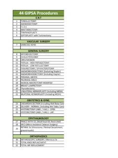
Transcription of ACROMIO-CLAVICULAR JOINT (AC JOINT) …
1 ACROMIO-CLAVICULAR JOINT (AC JOINT ) degeneration Anatomy of the AC JOINT : This is the small JOINT on the top of the shoulder. It connects the tip of the clavicle (collar bone) to the acromion (shoulder bone). It is held together by ligaments between the two bones as well as strong ligaments between the collar bone and the coracoid (a protrusion from the shoulder blade). ACROMIO-CLAVICULAR JOINT (AC JOINT ) pain is often referred to as AC degeneration . It occurs more in males than in females and is often related to sport, eg. rugby and others where repeated falls on the shoulder may lead to direct impact and damage of this small JOINT on top of the shoulder. It is not uncommon to see this condition in young sportsmen in the 20-30 age group. Other upper body sports like weight lifting is also known to cause this and one type of this condition is referred to as ( weight lifter s shoulder ).
2 Acute injuries to this JOINT most commonly lead to subluxations (dislocations) but not infrequently to damage ( tear) of the cartilage (meniscus) in the JOINT .. Anatomy: The ACROMIO-CLAVICULAR (AC) JOINT is situated on top of the shoulder and is the articulation between the clavicle (collar bone) and acromion (part of the shoulder blade or scapula). There is a meniscus (small cartilage cushion ) in the JOINT which absorbs the impact. If this meniscus is worn out either from repeated motion or injury the cartilage covering of the JOINT is worn away and contact of bone on bone of the JOINT surfaces results. A. Dislocation of the JOINT B. degeneration of AC JOINT degeneration of the ACJ with cavities in bony surfaces (cysts) The resulting inflammation may then lead to erosion of the ends of the bones of the JOINT , often seen as cysts on the bone ends (also referred to as moth-eaten appearance ) (shown below) Diagnosis: Symptoms: This condition usually causes pain on top of the shoulder; there is often some swelling in this area and tenderness with pressure.
3 The pain can refer medially to the trapezius and collar bone area. Sleeping on it usually causes pain and certain arm movements such as moving the arm across the body, behind the back and also high up overhead, elicit the pain: Exercises in the gym like bench press, push ups and overhead lifting motions can make the pain worse. An arthroscopic view of the degenerated AC JOINT . Right arrow: Meniscus, Left arrow: Denuded JOINT surface. Mechanism of pain in AC JOINT degeneration . The doctor would then do some tests: Tenderness would be felt with pressure on the top of the shoulder on the JOINT . Certain tests done by the doctor would then cause pain, especially forcing the arm of the patient across the chest. X-Rays: will show JOINT space narrowing and projections from the undersurface of the tip of the collar bone called osteophytes.
4 Cysts may also been seen in the tip of the bone. Ultra-sound: swelling with fluid in the AC JOINT will be evident. In our studies we have found this to be a reliable sign of an abnormal AC JOINT . Test: ACJ tenderness. To diagnose this condition accurately we inject local anaesthetic into the AC JOINT with ultrasound guidance to ensure that the needle is accurately placed inside the JOINT . If the pain disappears following the injection, this confirms the diagnosis beyond any doubt. The local anaesthetic may be mixed with a small amount of cortisone which may have a longer effect for pain relief. Unfortunately, as this is a mechanical problem, the relief lasts only a few days but such a response is at least highly diagnostic. Such a response is also an indicator that an operation to clear out the JOINT will be successful.
5 Management As a first line, conservative treatment is acceptable- there is never a rush to operate this condition as it does not cause any long-term problems. The only indication for any intervention is the degree of pain. Conservative measures would include anti-inflammatory medication, application of ice and physiotherapy. The above-mentioned injections may also lead to pain relief but is seldom curative Surgical treatment: We propose an arthroscopic operation The arthroscope is a small tube with a camera attached, and the surgeon can see a picture on a large TV screen and through another small hole instruments are passed to do the procedure: this means that the operation is done through two small holes 3-4 mm in size. There are hardly any scars visible after the operation. Overview of the procedure. Arthroscopic excision.
6 The first part of the operation is that all the torn material (cartilage, etc) is thoroughly removed. A small amount of bone is then shaved of each side of the JOINT as this bone is usually degenerated and sick . This allows for a small gap between the ends of the bone which then fills up with normal scar tissue and prevents any bone to bone contact. The JOINT then becomes pain-free as the new cushion develops between the ends of the bone Post Operatively: 1. No sling needs to be worn. 2. The patient is usually discharged on the same day following the surgery and is immediately allowed to do any activity that he is able to. 3. There may be some discomfort for a few days but there will be a gradual improvement and activities can be tailored to the pain with no other restriction. Arthroscopic decompression of the JOINT in progress (left) and a decompressed JOINT (right).
7 4. Therefore return to work, sports and everyday actions like driving, clerical and other duties simply depend on the discomfort from the operation and no harm will be done with such activities This is usually a very successful procedure and is often done on high level and professional athletes which enable them to return to their previous levels of sport. Function after surgery. Small scars after arthroscopic surgery