Transcription of Animal Structure and Function - San Diego Miramar College
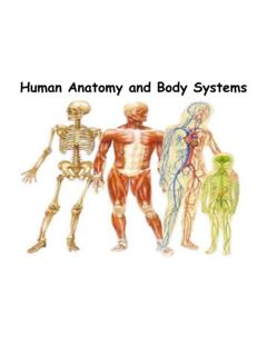
1 Animal Structure and Function (Outline) 1. Review levels of structural hierarchy of the living world 2. Define the terms anatomy and physiology. 3. Identify the four types of tissues in animals, their basic Structure and Function . 4. Learn the 4 types of epithelial cells with examples and their location and Function . 5. Learn the importance of connective tissue, the different types and their Function . Compare and contrast cartilage, bone, tendons, and ligaments . 6. Learn the basic Structure of muscle and the three different types and their Function . 7. Learn the Structure of nerves and their Function . 8. Define an organ and the organization of different tissues within. 9. Learn the 10 organs systems of the Animal body, their overall functions, and organs.
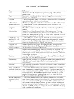
2 And contrast early bodies of early animals with the three -layered large complex Animal bodies. the importance of homeostasis for animals and review how warm blooded organisms maintain their body temperature. Atom Molecules Organelle Cell Tissue Organ Organ system Organism Population Community Ecosystem Bioshpere - Life is organized into a hierarchy of structural levels - Each level builds on the level below it (Emergence/ emergent properties) Anatomy & Physiology Rows of setae on a gecko s foot Spatulae coming from a single seta Function : Walking on walls and ceilings by Salamander & small lizards (Geckos) Structure : Hairs on toes (setae) are split into spatulae. Molecules on spatulae adhere to solid surfaces. Biological Theme: Structure fits Function in the Animal body Anatomy is the study of Structure Physiology studies how structures Function Flight Function depends on specific structures of wings, bone.
3 And pectoral muscle Palm Finger 2 Finger 3 Shaft Barb Barbule Hook Feather Structure Wrist Forearm Finger 1 Internal bone Structure Shaft Figure Structure in the living world including that of animals is organized in a series of hierarchical levels Muscle cell A Cellular level B Tissue level Muscle tissue C Organ level Heart E Organism level Many organ systems functioning together D Organ system level Circulatory system Figure E Structure in lab Function in lab Tissues are groups of many similar cells that perform the same specific Function Tissue types Epithelial tissue Connective Muscle Nervous Epithelial Tissue Structure : Closely packed sheets of cells Cover surfaces and line the cavities and tubes of internal organs Functions: Protection Exchange.
4 Secretion, absorption Excretion-waste products Sensation Flat Cube-shaped Column-shaped Single layer on a basement membrane (connective tissue) Multiple layers on a basement membrane (connective tissue) Basement membrane (extracellular matrix) Free surface of epithelium Cell nuclei A Simple squamous epithelium (lining the air sacs of the lung) B Simple cuboidal epithelium (forming a tube in the kidney) C Simple columnar epithelium (lining the intestine) D Stratified squamous epithelium (lining the esophagus) Layers of dead cells Rapidly dividing epithelial cells E Stratified squamous epithelium (human skin) Colorized SEM Figure E Underlying tissue Simple Epithelium Squamous mouth, blood vessels, heart, lungs and outer layers of the skin Cuboidal Glands and their ducts, and the lining of the kidney tubules Columnar lining of the stomach and intestines Some specialized for sensory reception: nose, ears and taste buds of the tongue oSome ciliated for directing flow oOther glandular producing and secreting.
5 Enzymes, hormones, milk, mucus, sweat, wax and saliva Stratified epithelium Keratinized top layer (tough)- skin Un-keratinized top layer- mouth cavity Epithelial tissue on the interior body surfaces is known as endothelium Connective Tissue Structure characterized by few cells in and large amount of extracellular non-living matrix secreted by its cells Liquid matrix (Blood) Semi-solid matrix (Tendons & others) Solid (Bone) Functions binds and supports other tissues Movement Many others Collagen osponge-like scaffold of a tensil protein Cartilage oSpecialized cells with extracellular matrix and proteins (collagen and elastin) Bone oliving and dead cells in the mineralized organic matrix ohardened by calcium phosphate and calcium carbonate deposits Ligaments oconnect bones to bone Tendons oconnect muscle to bone Cartilage- forming cells B Fibrous connective tissue (forming a tendon) Matrix D Cartilage (at the end of a bone) Central canal Matrix Bone- forming cells E Bone F Blood Elastic fibers Collagen fiber Cell Collagen fibers Cell nucleus White blood cells Red blood cell Plasma C Adipose tissue Fat droplets Figure F B Fibrous connective tissue (forming a tendon) A Loose connective tissue (under the skin)
6 Muscle Tissue Structure Fibers made of many fused cells that have contractile proteins and multiple nuclei Three types of muscles Skeletal: voluntary body movements Cardiac : pumps blood Smooth: involuntary moves the walls of internal hollow organs, such as the GI, arteries, bladder, uterus. Function Movement & mechanical work Unit of muscle contraction Muscle fiber Nucleus A Skeletal muscle Nucleus Muscle fiber Junction between two cells Muscle fiber Nucleus C Smooth muscle B Cardiac muscle Figure C Nervous Tissue Structure Neurons that make up the brain, spinal cord and peripheral nerves that branch throughout the body Branching neurons made of a cell body and have cell extensions: axon, and dendrites Function Communication network Transmit nerve signals rapidly to control body activities Cell body Nucleus Cell extensions An organ is made of several tissues that collectively perform specific functions Figure Small intestine (cut open) Lumen Epithelial tissue (columnar epithelium) Connective tissue Smooth muscle tissue (2 layers) Connective tissue Epithelial tissue Lumen Organ systems work together to perform life functions.
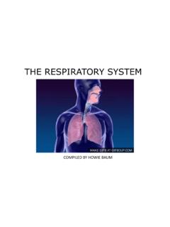
7 Each organ system has one or more functions Eleven organ systems: digestive Respiratory Circulatory Immune Excretory Endocrine Nervous Integumentary Skeletal Muscular Reproductive The digestive and respiratory systems Gather food and oxygen Gather oxygen Digest & absorb Send oxygen to heart Remove undigested food Remove carbon dioxide A digestive system Mouth Esophagus Liver Stomach Small intestine Large intestine Anus B Respiratory system Nasal cavity Larynx Trachea Bronchus Lung Figure , B The circulatory system and the lymphatic system Transports the food and oxygen collect and circulate liquid to and from tissues The immune system Protects the body from infection and cancer C Circulatory system Heart Blood vessels E Lymphatic system D Immune system Bone marrow Thymus Spleen Lymph nodes Lymph vessels Figure E C Lymphatic system The excretory system The endocrine and nervous systems Filters blood Control body functions Disposes of certain wastes F Excretory system Kidney Ureter Urinary bladder Urethra Pituitary gland Thymus Thyroid gland Te s t i s (male) Adrenal gland Pancreas G Endocrine system Ovary (female)
8 The integumentary system Skeletal and muscular systems Covers and protects the body Support and move the body I Integumentary system Hair Skin Nails K Muscular system Skeletal muscles Cartilage Bones J Skeletal system The Reproductive system Production of gametes Perpetuates the species Figure Female Vas deferens Penis Urethra Te s t i s Prostate gland Male Oviduct Ovary Uterus Vagina L Reproductive systems Figure The Primordial Embryo Animals exchange materials with their environment Structural adaptation include shape and size: Small with 2 layers for material exchange Large with increased surface area and specialized structures Small & simple body construction Diffusion Two cell layers Diffusion Mouth Gastrovascular cavity Figure Larger & complex animals specialized structures that increase surface area Exchange of materials between blood and body cells via the interstitial fluid Respiratory system Excretory system digestive system Circulatory system External environment Food Mouth Animal Body cells Interstitial fluid Anus Unabsorbed matter (feces) Metabolic waste products (urine)
9 Intestine Nutrients CO2 O2 Figure The respiratory system with its enormous internal surface area Figure Animals regulate their internal environment to achieve an internal steady state, homeostasis. Homeostatic mechanisms External environment Internal environment Small fluctuations Large fluctuations Figure Figure Homeostasis depends on negative feedback to keep internal variables fairly constant, with small fluctuations around set points Homeostasis: Internal body temperature of approximately 36 38 C Temperature rises above normal Temperature falls below normal Temperature decreases Temperature increases Thermostat shuts off warming mechanisms Blood vessels in skin constrict, minimizing heat loss Thermostat in brain activates warming mechanisms Skeletal muscles rapidly contract, causing shivering, which generates heat Thermostat in brain activates cooling mechanisms Sweat glands secrete sweat that evaporates, cooling body Blood vessels in skin dilate and heat escapes Thermostat shuts off cooling mechanisms Figure