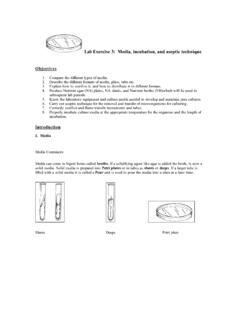
Transcription of Chromosomes, Karyotyping, and Abnormalities
1 1 Chromosomes, Karyotyping, and Abnormalities (Learning Objectives) Learn the components and parts of a metaphase chromosome . Define the terms karyotype, autosomaland sex chromosomes. Explain how many of each is present in a gamete and in a somatic cell. Explain the field of cytogeneticsand its uses for visualization of chromosomes, karyotypingfor prenatal and postnatal diagnoses, including the sources of cells used. Explain the differences between the three prenatal diagnosis techniques. Learn the terms used to describe the Abnormalities in chromosomal numbers: polyploidy, aneuploidy: trisomyand monosomy, and mosiacismand their causing mechanisms.
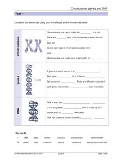
2 Learn the terms that describe the Abnormalities in chromosomal structure: deletions, duplications, translocations, and inversions. Learn the basics of the shorthand used to describe normal and abnormal karyotypes. Recognize the common autosomaland sex chromosome aneuploidies. 2 Portrait of a ChromosomeFigure of a ChromosomeA chromosome consists primarily of DNA and proteinChromosome differ in size and shapeEssential parts are:- Telomeres- Origins of replication sites - Centromere4 Portrait of a ChromosomeHeterochromatinis darkly staining- Consists mostly of repetitive DNAE uchromatinis lighter-staining - Contains most protein-encoding genesTelomeresare chromosome tips composed of many repeats of TTAGGG - Shorten with each cell division5 CentromeresThe largest constriction of the chromosome -attachment sites of spindle fibersDNA present at the centromere are specific repeated sequence 6 SubtelomeresThe region between the centromere and telomeres Consists of 8,000 to 300.
3 000 basesNear telomere the repeats are similar to the telomere sequence 7 Figure Chromosomes1882 Figure by German biologist Walther FlemmingNowMicrograph of actual stained human chromosomes9 CytogeneticsVariations in chromosomal structure occur as cells go through the cell cycleCytogeneticsis a technical field within genetics for visualization of chromosomal variationsExcess genetic material has milder effects on health than a deficitMost large-scale chromosomal Abnormalities disrupt or halt prenatal development10 These are sex chromosomesThe chromosome pairs 1 trough 22 are autosomeKaryotypeA visual display of metaphase chromosomes arranged by size and structure11 KaryotypeA visual display of chromosomes arranged by size and structure- Autosomes are numbered 1-22 by size- Sex chromosomes are X and Y Humans have 24 different chromosome types12 Figure PositionsPosition of centromeres varies between chromosomes At tip Telocentric Close to end Acrocentric Off-center Submetacentric At midpoint MetacentricFigure are useful1) Can confirm a clinical diagnosis2) Can reveal effects of environmental toxins3)
4 Can clarify evolutionary relationships15 Sources used for KaryotypingTissue is obtained from person- Fetal tissue: AmniocentesisChorionic villi samplingFetal cell sorting chromosome microarray analysis- Adult tissue: White blood cellsSkin-like cells from cheek swab Chromosomes are extractedThen stained with a combination of dyes and DNA probes16 Prenatal Diagnosis: AmniocentesisDetects about 1,000 of the more than 5,000 known chromosomal and biochemical problemsUltrasound is used to follow needle s movementFigure Diagnosis: Chorionic Villi Sampling Performed during 10-12thweek of pregnancy Provides earlier results than amniocentesis Limited detection of some metabolic problems Has greater risk of spontaneous abortionFigure Diagnosis.
5 Fetal Cell SortingFetal cells are distinguished from maternal cells by a fluorescence-activated cell sorter - Identifies cell-surface markersFigure new technique detects fetal mRNA in the bloodstream of the mother19 FISHF luorescence in situhybridization DNA probes labeled with fluorescing dye bind complementary DNAF igure dots correspond to three copies of chromosome 2120 chromosome AbnormalitiesA karyotype may be abnormal in two ways:1) In chromosome number2) In chromosome structureAbnormal chromosomes account for at least 50% of spontaneous abortionsDue to improved technology, more people are being diagnosed with chromosomal abnormalities21 Table Cell with extra set of chromosomes is polyploid Triploid(3N) cells have three sets of chromosomes- Produced in one of two main ways.
6 OFertilization of one egg by two spermoFusion of haploid and diploid gametes Triploids account for 17% of all spontaneous abortions and 3% of stillbirths and newborn deaths 24 TriploidyFigure A normal chromosomal number is euploid Cells with extra or missing chromosomes areaneuploid, gain or loss of a single chromosome Most autosomal aneuploids are spontaneously aborted Those that are born are more likely to have an extra chromosome (trisomy) rather than a missing one (monosomy)26 Nondisjunction The failure of chromosomes to separate normally during meiosis Produces gamete with an extra chromosome and another with one missing chromosome Nondisjunction during Meiosis I results in copies of both homologs in one gamete Nondisjunction during Meiosis II results in both sister chromatids in one gamete27 Nondisjunction at Meiosis IFigure at Meiosis IIFigure also arise during mitosis after the zygote formation.
7 Producing groups of somatic cells with the extra or missing chromosomes autosomalaneuploidy sex chromosome aneuploidyAn individual with two chromosomally-distinct cell populations is called a mosaicA mitotic non-disjunction event that occurs early in development can have serious effects on the health of the individual30 AutosomalTrisomiesMost autosomal aneuploids cease developing as embryos or fetusesMost frequently seen trisomies in newborns are those of chromosomes 21, 18, and 13- Carry fewer genes than other autosomes31 Table chromosome AneuploidyMissing one or having one or more additional copies the sex chromosomes Turner syndrome Triplo-X Klinefelter Syndrome XXYY Syndrome XYY Syndrome33 chromosome Structural AbnormalitiesFigure Structural Abnormalities Deletionsomissing a segment from a chromosome DuplicationsoPresence of an extra segment on a chromosome (Deletions and duplications often not inherited, arise de novo)
8 Translocationsotwo non-homologous chromosomes exchange segmentsoBalanced and unbalanced translocations InversionsoA chromosomal segment is flipped in orientation35 Chromosomal Shorthand36 Duplications in chromosome 15 Figure