Transcription of Biology 2108 Laboratory Exercises: Variation in …
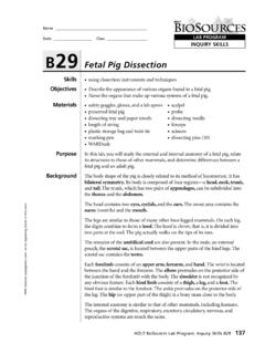
1 1 Biology 2108 Laboratory Exercises: Variation in Natural Systems Ed Bostick Don Davis Marcus C. Davis Joe Dirnberger Bill Ensign Ben Golden Lynelle Golden Paula Jackson Ron Matson Paul Pam Rhyne Gail Schiffer Heather Sutton Kennesaw State University Department of Biology and Physics Laboratory 5 Anatomy: fetal Pig dissection 2 OVERVIEW OF LAB Like cells and ecosystems, organisms are units composed of many parts that interact and function together. The organism as a whole is composed of organ systems, such as the nervous system, and the circulatory system. Organ systems are composed of organs (such as the brain and the heart), and these, in turn, are made up of tissues. The small intestine, for example, is made up of epithelial, connective, and muscle tissues arranged in concentric circles.
2 In this lab, you will dissect a fetal pig, examining the general anatomy of a vertebrate. In addition to the dissection , you will look up the functions of specific organs in order to understand how they work with one another. Learning skills is an important component of most lab courses. So far, for example, you have had practice in designing experiments, taking accurate measurements, and using statistics. In this lab you will learn how to dissect and how to use a microscope. Carefully read and follow the directions given. You will receive two grades for this lab. The first will be an evaluation of your dissection based on the thoroughness and correctness of your work. The second will be your score on a lab practical.
3 REMEMBER: You will be denied access to the Laboratory if you are not appropriately dressed. Shorts, short skirts and open-toed shoes are not allowed. You must wear safety glasses and gloves (provided in lab). Students may wish to purchase their own gloves and dissecting tools. Safety glasses will be provided. dissection OF THE fetal PIG dissection Tools scissors, forceps, blunt probe. The following is a list of structures which you must locate. You are also responsible for the function of structures with an asterisk. These functions can be found in your text and in the supplemental Laboratory manuals in the lab. Structure Function or Special Features A. External Anatomy head neck trunk appendages tail mouth lips cheeks external nares eyes eyelids auricle external acoustic meatus thorax abdomen umbilical cord prepuce orifice scrotum anus labia genital papillae vulva mammary papillae hooves wrist ankle elbow knee 3 Structure Function or Special Features B.
4 Internal Anatomy Digestive and Respiratory Systems masseter parotid gland epiglottis* teeth tongue papillae of tongue hard palate soft palate oral cavity nares (nasal cavities) nasal septum pharynx larynx* thymus* vocal cords trachea glottis thyroid* esophagus diaphragm* coelom pleural cavities lungs* pericardial sac parietal pleura visceral pleura parietal pericardium visceral pericardium visceral peritoneum mesenteries liver* umbilical vein* umbilical arteries* gall baldder* stomach* lesser curvature greater curvature greater omentum spleen* pyloric valve* duodenum* jejuno-ilium* pancreas* colon* caecum rectum* anus 4 Structure Function or Special Features C.
5 Heart and Great Vessels coronary arteries and veins* right and left atria* right and left ventricles* cranial and caudal vena cava* pulmonary trunk* pulmonary artery pulmonary vein aorta* D. Excretory System kidneys* adrenal gland* ureter* urinary bladder* urethra* E. Reproductive System scrotum* testes* inguinal canal epididymis* ductus deferens* spermatic cord glans penis prepuce ovaries* uterine tube* horn of the uterus body of the uterus* vagina vestibule 5 INTRODUCTION As you begin to dissect the fetal pig, you must keep in mind the purpose of a dissection . In a dissection , you expose important structures in order to see their relationship to each other.
6 It is, therefore, important not to destroy or remove organs and to accomplish the dissection so carefully that another student would be able to locate all of the structures in their appropriate location. You will only use scissors to make the initial incisions. Most of the rest of the dissection can be completed using the blunt probe and forceps. During the reading of the instructions, you will find several terms to be useful. They are defined below: superficial fascia - the loose connective tissue beneath the skin and surrounding organs. This material can be picked away with forceps. adipose tissue - fat beneath the skin right and left - the pig s right and left medial - the midline of the body or toward the midline of the body lateral - toward the sides of away from the midline of the body superficial - close to the surface of the body deep - further from the surface of the body anterior (cranial) - toward the head posterior (caudal)
7 - towards the tail ventral - belly surface dorsal - back surface distal - point of a structure, usually on a limb, farthest from the midline proximal - point of a structure closest to the midline 6 EXTERNAL ANATOMY Examine the body of the pig and note that it consists of 4 basic sections; the head, neck, trunk, and tail. Two pairs of appendages are attached to the trunk. Look at the head and locate the mouth surrounded by lips and cheeks which help the young to nurse. At the end of the snout are the nares, and cranial and lateral to them are the eyes with eyelids. Make a small incision in the medial corner of the eye and by pulling the eyelids apart, locate the nicitating membrane which helps to keep the eyeball clean.
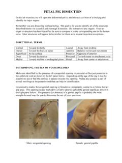
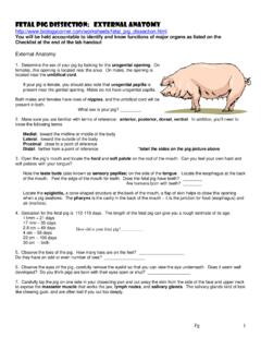
8 Lateral on the head opposite the auricle which helps to gather sound waves and the opening of the ear, the external acoustic meatus. The trunk of the pig consists of a cranial thorax, a caudal abdomen, and a dorsal lumbar region. On the ventral surface of the fetal pig, locate the umbilical cord which connects to the placenta. The male pig has a prepuce orifice of the penis and the scrotum both caudal to the umbilical cord. The female pig has a common opening to the urinary track and vagina located between the folds or labia. The labia come together ventrally to form the genital papilla. The external genitals of the female are called the vulva. In both sexes locate the end of the digestive track, the anus, and the five or six pairs of mammary papillae.
9 Observe the appendages attached to the trunk. The pig is called an ungulate because it walks on the tips of its toes. Notice the elongation of the feet and how the wrist and ankle are carried off the ground. They are easily confused with the elbow and knee. INTERNAL ANATOMY A. Digestive and Respiratory Systems Systems consist of organs which are grouped together to perform a specific function. The digestive system is responsible for the mechanical and chemical hydrolysis (digestion) of the food we eat and for the absorption of nutrients. Gases are exchanged by the respiratory system. The oxygen is needed by the body to help produce energy and the carbon dioxide is a waste product of this energy production.
10 Carefully remove the skin from one side of the head and neck in order to expose the salivary glands, often confused with the muscles. Glandular tissue has a lumpy appearance while muscle tissue can be recognized by parallel stripes caused by the arrangement of the muscle cells into bundles. You may first notice the large masseter muscle at the angle of the lower jaw. There are several different salivary glands but the one easiest to locate is the parotid gland located just caudal to the masseter muscle. Insert your scissors into the angle of the lips on the side of the head not yet dissected. Make an incision following the curvature of the tongue being careful not to cut into the roof of the mouth.