Transcription of ch 07 MANUAL PLATELET COUNT - Pearson …
1 MANUAL PLATELET COUNTP rincipleWhole blood is diluted with a 1% ammonium oxalate solution. The isotonic balance of thediluent is such that all erythrocytes are lysed while the leukocytes, platelets, and reticulocytesremain ,2 The standard dilution for PLATELET counts is 1:100. This dilution is prepared usingthe leukocyte/ PLATELET Unopette The dilution is mixed well and incubated to permit lysisof the erythrocytes. Following the incubation period, the dilution is mounted on ahemacytometer. The cells are allowed to settle and then are counted in a specific area of thehemacytometer chamber under the microscope. The number of platelets is calculated per L (x109/L) of and leukocyte/ PLATELET Unopette reservoirs; each containing mL of the following diluent:Ammonium gSorensen s phosphate buffer gQS with distilled water to 1 Unopette capillary pipets, 20 with cover dish with filter counter6.
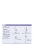
2 MicroscopeQuality ControlCommercial quality control materials with established control limits should be run frequency is determined by each laboratory's workload. For instance, quality controlmaterial may be run at the beginning of each eight-hour blood, anticoagulated with EDTA, or free-flowing capillary blood may be two leukocyte/ PLATELET Unopettes as follows:a. Using the protective shield on the capillary pipet, puncture the diaphragm asfollows:1)Place reservoir on a flat surface. Grasping the reservoir in one hand, takethe pipet assembly in the other hand and push the tip of the pipet shieldfirmly through the diaphragm in the neck of the reservoir, then Remove the shield from the pipet assembly with a twist and fill the capillary pipetwith whole blood.
3 Transfer the whole blood to reservoir as follows:1)Wipe excess blood from the outside of the capillary pipet, making certainthat no blood is removed from the capillary )Squeeze the reservoir slightly to force out some air. Maintain pressure onthe )Cover opening of overflow chamber of the pipet with your index fingerand seat the pipet securely in the reservoir )Release pressure on the reservoir. Then remove your finger from the pipetopening. Negative pressure will draw the blood into the )Squeeze the reservoir gently two or three times to rinse the capillary bore,forcing diluent into, but not out of, the overflow chamber, releasing pressureeach time to return the mixture to the )Place your index finger over the pipet opening and gently invert severaltimes to thoroughly mix the blood with diluent.
4 7)Let stand for 10 minutes to allow erythrocytes to the hemacytometer and cover glass by flooding them with 70% alcohol. Drythoroughly with gauze or tissue; do not allow the alcohol to dry on the sure to remove all lint. Place the cover glass in position over the ruled Following incubation, mix diluted blood thoroughly by inverting reservoir to resuspendcells. Charge hemacytometer as to dropper assembly by withdrawing the pipet from the reservoir andreseating it securely in its reverse the capillary bore by inverting the reservoir, gently squeeze the sides, anddiscard the first three or four the pipet tip on the edge of the ruled area of the counting charge the hemacytometer with diluted blood by gently squeezing thesides of the reservoir to expel the contents until the chamber is properly procedure to charge the other side of the hemacytometer with the firstUnopette the hemacytometer on moistened filter paper in a Petri dish, and allow tostand 10 minutes to permit the cells to this same procedure.
5 Charge a second hemacytometer with the second Unopette place the hemacytometer on the microscope stage. Perform cell COUNT asfollows:a. With the low-power (10x) objective, locate the ruled area and the center largesquare (1 mm2). Examine the entire center square for even distribution ofplatelets, then carefully switch to the high-dry-power (40x) phase objective forcounting platelets. With phase microscopy, platelets appear as round or are counted in the entire center large square (1 mm2) (Figure 7-7) asfollows:1) COUNT the platelets in the first row of squares going from left to right, thenfrom right to left in the second row; follow this pattern until all rows )Within each square, COUNT all platelets touching the top and left-handborders.
6 Do not COUNT any cells touching the bottom or right-hand )Use the fine adjustment knob to focus up and down to identify this counting procedure for the other side of the the counts for each center square. The difference between these twocounts should not exceed 10%.e. COUNT the platelets on the second hemacytometer following the above The calculation formula for hemacytometer cell counts determines the number of cellswithin 1 L (1 mm3) of blood (Figure 7-9). To make this determination, the total numberof cells counted must be corrected for the initial dilution of blood and the volume ofdiluted blood used. The standard dilution of blood for PLATELET counts is 1:100; thereforethe dilution factor is 100.
7 The volume of diluted blood used is based on the area anddepth of the counting area. The area counted is 2 mm2 and the depth is mm; thereforethe volume factor is mm3 .Total number of cells counted dilution factor 1/volume factor = cells/mm3 Cells/mm3 = cells/ L or cells/ L 103 L /L = cells x 109/LExample: 100 x 103 platelets/ L 103 L/L = 100 x 109 the PLATELET counts from the duplicate pipets and report result (x 109/L or /mm3).Reference Interval150-440 x 109 counts should be performed within three hours after the dilution has been coefficient of variation (CV) for the 95% confidence limits is + 22%.3. If blood is collected by a skin puncture, carefully remove the first drop of blood andcollect free-flowing blood for the PLATELET COUNT .
8 This will minimize the occurrence ofplatelet clumping and adhesion of platelets to the puncture clumps of platelets are seen in the hemacytometer, the procedure should be may be due to inadequate mixing of blood or to poor technique in obtaining theblood Wright's-stained peripheral blood smear should be examined and the PLATELET estimatedetermined to confirm the hemacytometer PLATELET COUNT . The PLATELET estimate shouldcorrelate with the PLATELET COUNT + 25%. If a discrepancy exists, the PLATELET COUNT andperipheral blood smear estimate should be acute leukemia, there is an increase of blast cells in the peripheral blood. Oftenfragments of cytoplasm about the size of platelets break off the blast cells.
9 Thesecytoplasmic fragments are called hyaline bodies. They are the same size and density asplatelets and may be counted as platelets by the hemacytometer or automated methods. Itis very important that all PLATELET counts be confirmed by slide examination so that a falseincrease of platelets (due to the counting of fragments) is not satellitism will result in falsely decreased PLATELET counts. With plateletsatellitism, platelets adhere to neutrophils when the blood sample is anticoagulated withEDTA and is not free to be counted. PLATELET satellitism can be corrected by redrawing theblood sample using sodium citrate as the anticoagulant. The resulting PLATELET COUNT mustbe multiplied by to account for the dilutional effect of the citrate second method for MANUAL PLATELET counts is the Rees Ecker method.
10 Using theerythrocyte diluting pipet, whole blood is diluted with a solution containing brilliantcresyl blue, which stains the platelets a light bluish color. The platelets are then countedusing a standard hemacytometer and bright field sources of error in hemacytometer cell counts are given in Web Table Unopette WBC/ PLATELET determination for MANUAL , : Becton, Dickinson, and Company; Brecher G, Cronkite EP. Morphology and enumeration of human blood platelets. J ApplPhysiol. 1950; 3 ' 1995-2003 by A Pearson Company Prentice-Hall, Inc. Legal Notic