Transcription of Chapter 20: The Cardiovascular System: The Heart
1 Chapter 20: The Cardiovascular system : The Heart Chapter Objectives ANATOMY OF THE Heart 1. Describe the location and orientation of the Heart within the thorax and mediastinal cavity. 2. Describe the layers of the pericardium and state each layer s function. 3. Examine the three layers of the Heart wall and know what type of tissue is found in each layer. 4. Know the names of the valves of the Heart . Know what type of valve each is and the exact location of each valve within the Heart . 5. Explain the basic function of the Heart valves. 6. Describe how the two atrioventricular valves work during a cycle of the Heart . 7. Describe the difference in structure of the semilunar valves compared with the atrioventricular valves and show how they work.
2 CARDIAC MUSCLE AND THE CARDIAC CONDUCTION system 8. Explain why the conduction system of the Heart is different in terms of electrical signaling. 9. Describe the sequence in which the cardiac action potential spreads through the conduction system . 10. Describe an action potential in cardiac contractile fibers in terms of depolarization, plateau, and repolarization. THE CARDIAC CYCLE 11. Define systole and diastole. 12. Explain how the passage of depolarization and repolarization through different areas of the Heart is related to specific components of the wave pattern of an electrocardiogram (ECG). 13. Demonstrate how the various components of the ECG correlate to the events occurring within the chambers of the Heart .
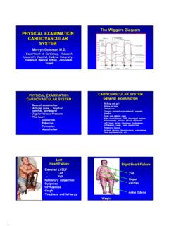
3 14. State which Heart sound is the result of the atrioventricular valves closing and which is the result of the semilunar valves closing. 15. Define End diastolic volume, End systolic volume and Stroke volume. 16. Describe what occurs to ventricular pressure during the following phases of the Cardiac cycle: isovolumetric relaxation, ventricular filling, and ventricular systole. CARDIAC OUTPUT 17. Define cardiac output (CO), describe the factors that affect it, and note changes with exercise in relation to cardiac reserve. 18. Define Preload, Contractility, and Afterload. 19. Explain that change in Heart rate is controlled by the CNS and hormones so as to influence cardiac output and blood pressure.
4 Chapter Lecture Notes Introduction The Heart is the pump that circulates the blood through an estimated 60,000 miles of blood vessels Heart pumps over 1 million gallons per year The Heart is situated between the lungs in the mediastinum with about two-thirds of its mass to the left of the midline (Fig ) The study of the normal Heart and diseases associated with it is known as cardiology Pericardium The Heart is enclosed and held in place by the pericardium. (Fig ) The pericardium consists of an outer fibrous pericardium and an inner serous pericardium (epicardium) Fibrous pericardium dense irregular CT protects and anchors the Heart , prevents overstretching The serous pericardium is a thin delicate membrane composed of a outer, parietal layer and an inner, visceral layer (epicardium)
5 Between the parietal and visceral layers is the pericardial cavity which is filled with pericardial fluid that reduces friction between the two membranes Pericarditis - inflammation of the pericardium Cardiac tamponade - bleeding into the pericardial cavity which compresses the Heart and is potentially lethal Layers of the Heart Wall The wall of the Heart has three layers: epicardium, myocardium, and endocardium (Fig ) The epicardium (visceral pericardium) consists of simple squamous epithelium and connective tissue The myocardium is composed of cardiac muscle and is the bulk of the Heart Cardiac muscle fibers swirl diagonally around the Heart in interlacing bundles Myocarditis - inflammation of the myocardium The endocardium consists of simple squamous epithelium and connective tissue chamber lining & valves Endocarditis - inflammation of the endocardium.
6 It usually involves the Heart valves. Heart Valves The Heart has two types of valves (Fig & ) Atrioventricular valves (AV) Tricuspid valve between right atrium and right ventricle has three cusps composed of dense CT covered by endocardium Bicuspid valve - between left atrium and left ventricle has two cusps Pneumonic - LAMB (Left Atrioventricular, Mitral, or Bicuspid valve) Semilunar valves (SL) Pulmonary semilunar valve - blood flows from right ventricle into pulmonary trunk Aortic semilunar valve - blood flows from left ventricle into the ascending aorta Heart Valve Function Valves open and close in response to pressure changes as the Heart contracts and relaxes AV valves open and allow blood to flow from atria into ventricles when ventricular pressure is lower than atrial pressure (Fig )
7 Ventricles are relaxed chordae tendineae are slack papillary muscles are relaxed AV valves close preventing backflow of blood into atria ventricles are contracted increased blood pressure in ventricles push valve cusps closed chordae tendinae are pulled taut papillary muscles contract to pull cords and prevent cusps from everting Semilunar valves open with ventricular contraction allow blood to flow into pulmonary trunk and aorta Semilunar valves close with ventricular relaxation prevents blood from returning to ventricles blood fills valve cusps and tightly closes the SL valves Conduction system of Heart Cardiac muscle cells are autorhythmic cells because they are self-excitable.
8 They repeatedly generate spontaneous action potentials that then trigger Heart contractions. (Fig ) The autonomic nervous system and hormones, such as epinephrine, do modify the heartbeat (in terms of rate and strength of contraction), but they do not establish the fundamental rhythm caffeine & nicotine increase activity Sinoatrial (SA) node Autorhythmic cluster of cells in wall of right atrium SA node generates an Action potential spontaneously 90-100 times per minute begins Heart activity that spreads through the intercalated discs to all cardiac muscle cells in both atria excitation spreads to AV node Atrioventricular (AV) node in atrial septum AV node fires at 40-50 times per minute transmits signal to bundle of His Atrioventricular (AV)
9 Bundle - bundle of His the connection between atria and ventricles divides into bundle branches Right and left bundle branches - large diameter fibers that conduct signals quickly in the interventricular septum Purkinje fibers in trabeculae carneae, papillary muscles and the myocardium SA node sets pace since it is the fastest In 50 msec excitation spreads through both atria and down to AV node 100 msec delay at AV node due to smaller diameter fibers - allows atria to fully contract filling ventricles before ventricles contract In 50 msec excitation spreads through both ventricles simultaneously Action Potential and Contraction of Contractile Fibers An Action potential in a ventricular contractile fiber is characterized by rapid depolarization, plateau, and repolarization (Fig )
10 Depolarization Cardiac cell resting membrane potential is -90mv excitation spreads through gap junctions in the intercalated discs fast Na+ channels open for rapid depolarization Plateau phase 250 msec (only 1msec in neuron) slow Ca+2 channels open, let Ca+2 enter from outside cell and from storage in sarcoplasmic reticulum, while most K+ channels remain closed Ca+2 binds to troponin to allow for actin-myosin cross-bridge formation & tension development Repolarization Ca+2 channels close and K+ channels open & -90mv is restored as potassium leaves the cell Refractory period - the time interval when a second contraction cannot be triggered very long so Heart can fill longer in a cardiac muscle fiber than the contraction itself.