Transcription of Chapter 23: The Respiratory System
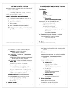
1 Chapter 23: The Respiratory SystemI. The Respiratory System : An Introduction, p. 814 Objectives:1. Describe the primary functions of the Respiratory Explain how the delicate Respiratory exchange surfaces are protected frompathogens, debris, and other hazards. Our cells produce energy for maintenance, growth, defense and division throughmechanisms which require oxygen and produce carbon dioxide. We obtainoxygen from the air, which diffuses across delicate exchange surfaces on ourlungs. Oxygen is carried to the cells by our cardiovascular System , which alsoreturns carbon dioxide to the of the Respiratory System , p.
2 814 The Respiratory System has 5 basic functions:1. Providing an extensive surface area for gas exchange between air andcirculating Moving air to and from the exchange surfaces of the Protecting Respiratory surfaces from the outside Producing Participating in the olfactory of the Respiratory System , p. 814 Figure 23-1 The Respiratory System is divided into the upper Respiratory System , above thelarynx, and the lower Respiratory System , from the larynx down. The Respiratory tract consists of a conducting portion (from the nasal cavity to theterminal bronchioles) and a Respiratory portion (the Respiratory bronchioles andalveoli).
3 Alveoli are the air-filled pockets within the lungs where all gas exchange 23-2 The Respiratory Epithelium For gases to exchange efficiently, the walls of the alveoli must be very thin, andthe surface area must be very great. The distance from the inside of an alveolus toand alveolar capillary is less than 1 micrometer. The surface area within the lungsis about 35 times the surface area of the body. The Respiratory mucosa, consisting of an epithelial layer and an areolar layer,lines the conducting portion of the Respiratory System . In the upper Respiratory System , trachea and bronchi: The lamina propria,underlying the areolar tissue, contains mucous glands that secrete onto theepithelial surface.
4 In the conducting portion of the lower Respiratory System : The lamina propriacontains smooth muscle cells that encircle the lumen of the bronchioles. The structure of the Respiratory epithelium changes along the Respiratory exchange surfaces of the alveoli are lined with alveolar epithelium: a verydelicate, simple squamous epithelium containing scattered, specialized cells. The Respiratory defense System consists of a series of filtration mechanisms thatremove particles and pathogens:- Goblet cells and mucous glands produce mucus that bathes Cilia sweep debris trapped in mucus toward the pharynx (mucusescalator).
5 - Filtration in the nasal cavity removes large Small particles that reach the lungs can be engulfed by The Upper Respiratory System , p. 817 Objective:1. Identify the organs of the upper Respiratory System and describe their 23-3 The Upper Respiratory SystemThe Nose and Nasal Cavity, p. 817 Air enters the Respiratory System through the nostrils or external nares into thenasal vestibule, which contains the first particle filtration System : nasal hairs. The nasal septum divides the nasal cavity into left and right. Mucous secretions from the paranasal sinus and tears keep the nasal cavity moistand clean.
6 The superior portion of the nasal cavity is the olfactory region whichprovides the sense of smell. Air flows from the vestibule to the internal nares through the superior, middle andinferior meatuses. The meatuses are constricted passageways that produce airturbulence, giving incoming air time to warm and humidify, and particles to betrapped. The hard palate forms the floor of the nasal cavity and separates it from the oralcavity. The soft palate extends posterior to the hard palate, dividing the superiornasopharynx form the rest of the pharynx. The nasal cavity opens into thenasopharynx through the internal nares.
7 The nasal mucosa prepares inhaled air for arrival at the lower Respiratory organs(warming and humidifying). Breathing through your mouth bypasses thisimportant Pharynx, p. 819 The pharynx is a chamber shared by the digestive and Respiratory systems,extending from the internal nares to the entrances to the larynx and esophagus. The pharynx is divided into the nasopharynx, the oropharynx, and thelaryngopharynx. The nasopharynx is the superior portion of the pharynx. It contains the pharyngealtonsils and openings to the left and right auditory tubes. The oropharynx, the middle portion of the pharynx, communicates with the oralcavity.
8 The laryngopharynx, the inferior portion of the pharynx, extends from the hyoidbone to the entrance to the larynx and The Larynx, p. 819 Objective:1. Describe the structure of the larynx and discuss its role in normal breathing andin production of sound. Air from the pharynx enters the larynx, a cartilaginous structure that surrounds 23-4 Anatomy of the LarynxCartilages and Ligaments of the Larynx, p. 819 Three large, unpaired cartilages form the larynx:1. The thyroid cartilage (Adam s apple): i. hyaline cartilage ii. forms anterior and lateral walls of larynx iii. ligaments attach to hyoid bone, epiglottis, laryngeal cartilages2.
9 The cricoid cartilage: iv. hyaline cartilage v. form posterior portion of larynx vi. ligaments attach to first tracheal cartilage vii. articulates with arytenoid cartilages3. The epiglottis: viii. elastic cartilage ix. ligaments attach to thyroid cartilage and hyoid bone The thyroid and cricoid cartilages surround and protect the glottis and theentrance to the trachea. During swallowing, the larynx is elevated and theepiglottis folds back over the glottis, preventing entry of food and liquids into therespiratory tract. The larynx also contains 3 pairs of smaller hyaline cartilages:1. the arytenoid cartilages2.
10 The corniculate cartilages3. the cuneiform cartilagesFigure 23-5 The Glottis The corniculate and arytenoid cartilages function in the opening and closing of theglottis and the production of sound. The vestibular ligaments and the vocal ligaments extend between the thyroidcartilage and the arytenoid cartilages, and are covered by folds of laryngealepithelium that project into the glottis. The vestibular ligaments lie within thevestibular folds, which protect the delicate vocal Production, p. 821 Air passing through the glottis vibrates the vocal folds and produces sound sound produced is varied by tension on the vocal folds, and on voluntarymuscles that position the arytenoid cartilage relative to the thyroid cartilage.