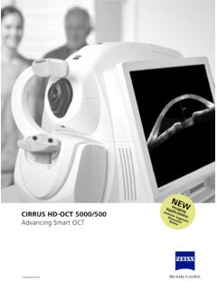
Transcription of Cirrus HD-OCT - ZEISS
1 Cirrus HD-OCTC ertainty in seconds. Certainty for know you ll love a both your patients and your practice in mind, Carl ZEISS Meditec, the global leader in OCT, developed Cirrus HD-OCT . Not only does it supply you with bar-setting imagery, it delivers detailed diagnostic and change analyses you can rely on time and again. Along with its small footprint and fast capture speeds, Cirrus is designed to improve workflow efficiency while helping you deliver better care to your s time to see what you ve been missing. It s time for CERTAINTY MAdE BY CARl ZEIssSuperior analysisWith high-density cube data and proven segmentation, Cirrus delivers a diagnostic analysis you can imagery With legendary ZEISS optics and Cirrus , you ll experience brilliant, detail-rich visuals to help you diagnose and care for your uses Selective Pixel Profiling to optimize each pixel in its HD Raster Scans.
2 It produces imagery that goes beyond mere image-averaging. It s a difference you need to see to Subfield Macular Thickness Repeatability Standard DeviationNo mMacular mDiabetic mVRI mSource: 510(k) Summary: Cirrus HD-OCT with Retinal Nerve Fiber Layer and Macular Normative Databases, Operating Characteristic Curves, Normal vs. Glaucomatous EyesParameterMildModerate to severeAverage RNFL Cup-to-Disc : Mwanza et al. Ability of Cirrus HD-OCT optic nerve head parameters to discriminate normal from glaucomatous eyes. Ophthalmology. 2011;118(2):241 5 Line Raster3D VisualizationLSO Fundus Image with ILM SlabAdvanced Visualization with ILM SlabEpiretinal MembraneHD 5 Line RasterPostsurgical Pseudophakic Cystoid Macular EdemaMacular Thickness MapLSO Fundus Image with Raster Line Macular Thickness MapILM Segmentation MapLSO Fundus Image with Macular Thickness Map3D VisualizationAdvanced Visualization with RPE Fit SlabAge-Related Macular DegenerationHD 5 Line RasterRPE Segmentation MapMacular Thickness MapCirrus gives you the ability to view pathologies from multiple vantage points and with a range of at-a-glance visualization formats, you ll be able to better assess your patient s condition and determine the appropriate course of pathology.
3 Multiple views. 45 Discover the power of the Cirrus offers unsurpassed OCT technology. Capturing a tightly packed, detail-rich cube of data in just seconds, it allows you to both visualize and analyze your patient s condition. Because the cube is populated with such high-density data, you can explore pathologies without requiring additional scan patterns. Scan with greater granularity Closely spaced B-scans within the cube ensure that even small areas of pathology are captured and easily viewable, unlike scans that are spaced further apart, which may miss the central fovea or nearby subtle defects. Enhance your analysis Millions of data points from the cube are fed into ZEISS proprietary algorithms for accurate segmentation, reproducible measurements and registration for change you can excellence Carl ZEISS Meditec and its research collaborators have developed advanced algorithms to measure and display , accurate centering of the measurement FoveaFinder and AutoCenter technologies ensure that measurements are made in the correct locations, taking the pressure off the operator to center the scans registration to track change Cirrus data cubes are automatically registered with data from prior visits, allowing for point-to-point data Diversified normative databases for ONH, RNFL and macular thickness facilitate at-a-glance assessments.
4 Generating a comprehensive cube of data is only the beginning. Cirrus gives you the ability to see beyond the scan and transform information into insight, becoming an indispensable part of your day-to-day clinical decision-making PatternData Points Per A-ScanTotal Data PointsSpacing Between LinesCapture Time512 x 1281024> 67 million47 s200 x 2001024> 40 million30 s67 FoveaFinder With FoveaFinder , Cirrus automatically and accurately locates the fovea, centers the ETDRS measurement grid and presents you with the B-scan through the fovea by your diagnostic certainty Cirrus enables rapid, careful assessment of the retina. By utilizing precise macular thickness analyses, providing detailed ILM and RPE layer maps and putting more than 100 B-scans at your disposal, Cirrus provides the framework to assess your patient s retinal subtle macular changeCirrus data cubes are automatically registered with data from prior visits after the scan is acquired.
5 This enables side-by-side visualization of the same location on the retina for each visit. Cirrus compares measurements from the current and prior visits to provide a thickness change map and helps you determine next steps for your RETINA dIAGNOsTICs MAdE BY CARl ZEIss89 Guided Progression Analysis (GPA ) compares RNFL thickness measurements from data cubes obtained during different visits and allows you to determine if statistically significant change has occurred over HFA- Cirrus Combined Report, available exclusively with ZEISS FORUM , summarizes patient structure and function information in a single display. AutoCenter After the scan is acquired, Cirrus automatically centers the measurement circle around the disc. The placement is not and track RNFL and ONH for glaucoma managementWith Cirrus , all traditional RNFL measurements based on the mm circle are present; however, Cirrus enables you to see past the circle-based assessments.
6 Spotting wedge defects and other patterns of loss is simplified with Deviation Maps, which show comparisons to normative data for each superpixel in the 6 x 6 mm Cirrus Optic Nerve Head analysis provides automated identification of the optic disc and cup boundaries. The analysis is generated using the dense data in the Optic Disc 200 x 200 data cube in tandem with a proprietary ZEISS algorithm. This algorithm precisely measures the neuroretinal rim while accounting for tilted discs, disruptions to the RPE and other challenging GlAUCOMA MANAGEMENT MAdE BY CARl ZEIss1013 For videos, presentations, recent clinical literature and updated product information, visit: represents the culmination of decades of patents, prototypes and progress. ZEISS is committed to delivering the excellence in installation, training and ongoing support you expect from the market new diagnostic needs emerge and new therapies are developed, innovation continues with Cirrus .
7 In addition, recognizing the modern electronic workplace, Cirrus integrates seamlessly into EMRs and with FORUM , our advanced data management solution for simplifying, centralizing and viewing the vast amounts of clinical data generated by ophthalmic instruments. There s only one OCT that promises you Certainty in Seconds, Certainty for Years. There s only one the industry leader in OCT, Cirrus is the best-selling spectral domain OCT in the your diagnostic insightCirrus offers anterior segment imaging of the angle and cornea and the ability to measure central cornea thickness. Narrow Angle VisualizationDescemet DetachmentMicrostriaeCentral Corneal Thickness MeasurementBullous Keratopathy with BCLC ataract IncisionIEK ZigzagKeratic PrecipitatesPRK Scar// ANTERIOR sEGMENT IMAGING MAdE BY CARl ZEIssSouth KoreaCarl ZEISS Co. 121-828 Mapo-gu141-1, Sangsu-dong2F, BR Elitel KoreaPhone: +82 2 3140 ZEISS Meditec Iberia de Poniente, 15 Tres Cantos28760 MadridSpainPhone: +34 91 203 37 ZEISS ABTegeluddsvaegen 7610254 StockholmSwedenPhone: +46 84 59 25 ZEISS AGFeldbachstrasse 818714 FeldbachSwitzerlandPhone: +41 55 254 ZEISS ThailandFloor 8, Thosapol Land Building 2230 Ratchadapisek RoadHuaykwang, Bangkok 10310 ThailandPhone: +66 2 2 74 06 KingdomCarl ZEISS Woodfield RoadWelwyn Garden CityHertfordshire, AL7 1 JQUnited KingdomPhone: +44 1707 States of AmericaCarl ZEISS Meditec, Hacienda DriveDublin, CA 94568 USAP hone: +1 925 557 local contact:ArgentinaCarl ZEISS Argentina Nahuel Huapi 4015 / 25C1430 BCO Buenos AiresArgentinaPhone: +54 11 45 45 66 ZEISS Pty.
8 13, 2 Eden Park DriveNorth Ryde, New South Wales 2113 AustraliaPhone: +61 2 9020 ZEISS GmbHLaxenburger Str. 21100 ViennaAustriaPhone: +43 1 79 51 ZEISS NV-SAIkaroslaan 491930 ZaventemBelgiumPhone: +32 2 719 39 ZEISS do Brasil Na oes Unidas, 21711 CEP04795-100 S o PauloBrazilPhone: +55 11 5693 ZEISS Canada Valleybrook DriveToronto, ON M3B 2S6 CanadaPhone: +1 800 387 ZEISS Shanghai Co. , Ke Yuan Building11 Ri Yin Nan RoadWaigaoqiao Free Trade Zone2005 Yang Gao Bei RoadShanghai 200131 ChinaPhone: +86 21 5048 17 RepublicCarl ZEISS spol. 14/3201150 00 Prague 5 Czech RepublicPhone: +420 233 101 ZEISS Meditec France SAS60, route de Sartrouville78230 Le PecqFrancePhone: +33 1 34 80 21 ZEISS Meditec VG mbHCarl- ZEISS -Strasse 2273446 OberkochenGermanyPhone: +49 7364 20 Ophthalmology:Phone: +49 800 470 50 KongCarl ZEISS Far East Co. 11-12, 25/FTower 2, Ever Gain PlazaNo.
9 88 Container Port RoadKwai ChungHong KongPhone: +852 2332 ZEISS India Pvt. , Kensington RoadUlsoorBangalore 560 008 IndiaPhone: +91 80 2557 88 ZEISS delle Industrie 2020020 Arese (Milan)ItalyPhone: +39 02 93773 ZEISS Meditec Japan Co. KuTokyo 160-000322 Honchio-ChoJapanOphthalmic instruments:Phone: +81 3 33 55 instruments:Phone: +81 3 33 55 ZEISS Sdn 2, Jalan 243/51 A46100 Petaling JayaSelangor Darul EhsanMalaysiaPhone: +60 3 7877 50 ZEISS de M xico de Miguel Angel de Quevedo 49604010 Mexico CityMexicoPhone: +52 55 59 99 ZEISS 300 Postbus 3103364 DL SliedrechtNetherlandsPhone: +31 184 43 34 ZealandCarl ZEISS ( ) Paramount Box 121 - 1001 Henderson, Auckland 0650 New ZealandPhone: +64 9 838 ZEISS sp. Z Lopuszanska 3202-220 WarsawPolandPhone: +48 22 858 ZEISS Ptd. Kaki Bukit PlaceSingapore 415926 SingaporePhone: +65 6741 AfricaCarl ZEISS (Pty.)
10 Oak AvenueFerndaleRandburg 2194 South AfricaPhone: +27 11 886 000000-1968-512 Rev B The contents of the brochure may differ from the current status of approval of the product in your country. Please contact your regional representative for more information. Subject to change in design and scope of delivery and as a result of ongoing technical development. Cirrus , FORUM, Selective Pixel Profiling, Advanced Visualization, Guided Progression Analysis, AutoCenter, FoveaFinder and Certainty in Seconds, Certainty in Years are either trademarks or registered trademarks of Carl ZEISS Meditec, Inc. in the United States and/or other countries. 2011 by Carl ZEISS Meditec, Inc. All copyrights reserved. 1011 5 MCarl ZEISS Meditec, Hacienda DriveDublin, CA ZEISS Meditec AGGoeschwitzer Str. 51-5207745 JenaGermany