Transcription of Comparative anatomy and phylogeny of the cloacae of ...
1 JOURNAL OF MORPHOLOGY 212~305-322 (1992) Comparative anatomy and phylogeny of the cloacae of Salamanders (Amphibia: Caudata). Vi. Ambystomatidae and Dicamptodontidae DAVID M. SEVER Department of Biology, Saint Mary's College, Notre Dame, Indiana 46556 ABSTRACT Histology of the cloacae of Rhyacotriton olympicus and represen- tative species from the genera Ambystoma and Dicamptodon was examined by light microscopy. Females of Ambystoma possess sperm storage glands, the spermathecae , as well as ventral glands and dorsal glands, both of uncertain function. Females of Ambystoma examined from the subgenus Linguaelapsus differ from those in the subgenus Ambystoma by possessing more extensive ventral gland clusters and a shorter cloacal tube. Females of Dicamptodon possess spermathecae and ventral glands, but differ in cloacal conformation from females of Ambystoma and lack the dorsal glands. Females of R. olympi- cus possess more extensive epidermal lining in the cloaca than that found in females of Ambystoma and Dicarnptodon, and the only glands present are spermathecae , which cluster around a tube in the dorsal roof.
2 Males of Ambystoma, Dicamptodon, and R. olympicus possess five types of cloacal glands (dorsal pelvic glands, lateral pelvic glands, anterior ventral glands, posterior ventral glands, and Kingsbury's glands) that function in spermato- phore formation, and vent glands that may produce a courtship pheromone. In Ambystoma and Dicamptodon, vent glands secrete along the medial borders of the cloacal orifice. Males of A. opacum and A. talpoideum differ from males of other species examined from the two genera by possessing more extensive vent glands. Males of R. olympicus possess unique vent glands in which tubules secrete onto the surface of vent lobes lateral to the posterior end of the cloacal orifice, and distal ends of the glands pass anteriorly, superficial to the fascia enclosing the other cloacal glands. The results from analysis of cloacal anatomy support other data indicating that Ambystoma and Dicamptodon are sister groups, and that Rhyacotriton olympicus is not closely related to either of the other two genera and merits placement in a separate family.
3 O 1992 Wiley-Liss, Inc. This paper reviews existing literature and presents additional descriptions of cloacal anatomy for the North American salamander families Ambystomatidae, containing the gen- era Ambystoma (27 species) and Rhyacosire- don (4 species), and Dicamptodontidae, con- sisting of Dicamptodon (4 species) and monotypic Rhyacotriton (Frost, '85). descrip - tions exist for the male cloacal anatomy of Dicamptodon tenebrosus and Rhyacotriton olympicus (Sever, '88a) and for eight species of Ambystoma (Sever, '81, '87, '88a; Sever et al., '89; Licht and Sever, '91). Descriptions of female cloacal anatomy , however, are limited to brief reports on A, tremblayi and R. olym- picus (Sever, '87) and more detailed studies on A. maculatum (Kingsbury, 1895) and A. gracile (Licht and Sever, '91). Duellman and Trueb ('86) illustrate the external appear- ance of the cloacal region of male and female A. jeffersonianum in breeding condition, and the relationships of the cloacal cavities to urogenital and digestive structures of sala- manders are shown by Sever ('92b).
4 In the present paper, the female cloacal anatomy of three species of Dicamptodon and seven spe- cies of Ambystoma is described for the first time. This paper is the sixth in a series on the Comparative anatomy and evolution of the cloacal region of salamanders (Sever, '91a,b; '92a-c). MATERIALS AND METHODS Snout-vent length (SVL) refers to the dis- tance from the tip of the snout to the poste- rior edge of the vent. Cloacal anatomy was o 1992 WILEY-LISS, INC. 306 SEVER examined following excision of the cloacal region and its histological preparation for light microscopy. Specimens examined (Ap- pendix I) were initially preserved in 10% neu- tral-buffered formalin, and were stored in 60% isopropanol or 65% ethanol before re- moval of cloacal tissue. Cloacal tissue was rinsed in water, dehy- drated in ethanol, cleared in toluene or Histo- sol (National Diagnostics, Inc., Manville, New Jersey), and embedded in paraffin; 10-pm sections were then cut with a rotary micro- tome.
5 Some sections from each species were stained with hematoxylin-eosin (for general cytology), and others were stained or treated with Mallory's triple stain (connective tis- sue), ninhydrin Schiff/fast green (proteins), periodic acid-Schiff s (PAS) reagentlfast green FCF (neutral carbohydrates), and/or alcian blue/nuclear fast red at pH , or toluidine blue (acid mucopolysaccharides). Staining procedures followed Humason ('79). Three-dimensional reconstructions were performed on transverse sections through the cloacae of a male Ambystoma laterale, and male and female A. opacum, Dicampto- don copei, and Rhyacotriton olympicus. These reconstructions were done using PC3D soft- ware (Jandel Scientific, Corte Madera, CAI and a Jandel digitizing tablet with a Zenith ZF-248 microcomputer. Every 4th to 15th section was digitized for image reconstruc- tion following procedures outlined by Sever Sever ('91a) used a ratio of cloacal tube length divided by total cloacal length (CTL/ TCL) to express the relative size of the cloa- cal tube.)
6 The cloacal tube is cylindrical and extends caudally to the cloacal chamber, a cavity dorsal to the vent. To determine lengths, the number of transverse sections of the cloacal tube and cloacal chamber was counted. The section immediately posterior to the junction of the Wolffian ducts with the hindgut was considered the most anterior section of the cloacal tube. Dicamptodon copei normally is an obligate paedomorph, and D. aterrimus and D. tene- brosus are facultative paedomorphs (Nuss- baum, '70, '83; Good, '89). Dicamptodon ater- rimus and D. tenebrosus prior to 1989 were considered conspecific with D. ensatus, a spe- cies now restricted to parts of northern Cali- fornia (Good, '89) and not examined for the present study. Maturity of branchiate fe- males was determined by large size of ovar- ian follicles, convolution of the oviducts, ('91a). and/or presence of sperm in the spermathe- cae. Stebbins ('54) reported that mature ova of D.
7 Ensatus are 5-6 mm in diameter, and Nussbaum ('85, '87) reported mean (ovipos- ited) ova sizes of mm for D. copei and mm or D. ensatus. In one Dicamptodon copei (UMMZ , mm SVL), ovarian follicles are only mm in diameter, but ovi- ducts are convoluted, and some sperm are present in the spermathecae ; thus this speci- men is mature. The other D. copei (UMMZ 134961) had the ovaries removed and lacks sperm in the spermathecae , but it is consid- ered mature because the oviducts are convo- luted and the specimen is even larger ( mm SVL) than other mature specimens. The only Dicamptodon aterrimus exam- ined (UMMZ 134671, 135 mm SVL) is meta- morphosed, has large ovarian follicles ( mm in diameter), and is thus considered mature; however, it has not recently mated since the spermathecae lack sperm. Of the three D. tenebrosus used in the present study, only one is metamorphosed and definitely mature. This individual (UMMZ 134955,279 mm SVL) has ovarian follicles about 5 mm in diameter and some sperm in the spermathe- cae.
8 Neither branchiate D. tenebrosus exam- ined possesses spermathecal sperm; both were collected September 13, 1975. One (UMMZ , 130 mm SVL) has ovarian follicles mm in diameter and is probably immature, while the other (UMMZ , 141 mm SVL) has ovarian follicles mm in diameter and may be mature. RESULTS Female cloacal anatomy Ambystomatidae Reconstructions based on serial sections through the cloaca of a female Ambystoma opacum are shown in Figure 1. Histological sections through cloacae are illustrated for A. opacum, A. gracile, A. maculatum, and A. tremblayi in Figure 2, and or A. annulatum and A. barbouri in Figure 3. The three types of glands Kingsbury (1895) reported for A. maculatum occur in all species. Interspecific variation occurs in cloacal conformation and anatomy of the ventral gland. The cloacal tube is present in all specimens examined except that of Ambystoma annulatum, in which the Wolffian ducts do not join until the anterior end of the cloacal tube, so that a definite Cloacal conformation.
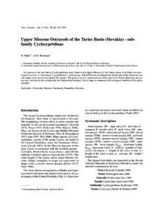
9 SALAMANDER cloacae 307 A C Fig. 1. Three-dimensional reconstructions of the clo- aca of a female Ambystoma opacum (UMMZ 187373). Right lateral view with sections rotated 25 clockwise. A: Walls of the cloacal cavities. B: spermathecae . C: Ante- rior ventral gland. D: Dorsal gland. The distance between cloacal tube is not present (Fig. 3A). In the other forms, the CTL/TCL quotient ranges from (A. texanum) to (A. laterale). In the species with a cloacal tube, the ante- rior half is narrowed dorsally and widened ventrally. The pseudostratified epithelium is formed into thick longitudinal folds or rugae (Fig. 2A) that are ciliated. At the midpoint of the cloacal tube, the dorsal half widens. In the posterior half of the cloacal tube, a pair of large folds appears in the lateral walls, form- ing a central groove between them (Figs. lA, 2A). Cilia occur on the epithelium of the cloacal walls lateral to the folds but not on the lining of the folds or the central groove.
10 Sections of the cloacal walls represents approximately 120 pm. Av, anterior ventral glands; Cc, cloacal chamber; Cg, central groove; Ct, cloacal tube; Dg, dorsal glands; St, spermathecae . Posteriorly, the large dorsolateral folds join medially, obliterating the central groove. In Ambystoma barbouri, A. jeffersonianum, A. laterale, A. opacum, A. platineum, and A. tremblayi, obliteration of the central groove occurs at the anterior end of the cloacal cham- ber (Figs. IA, 2B,C). In A. gracile, A. macula- tum, A. talpoideum, and A. tigrinum, the central groove ends prior to the claacal cham- ber, and the posterior fourth of the cloacal tube is a flattened slit (Fig. 2D). Since the cloacal tube is absent in A. annulatum, the central groove occurs in the anterior end of the cloacal chamber (Fig. 3A). The apex of the central groove is slanted posteriorly, so a 308 SEVER Fig. 2. Transverse sections through the cloacae of female Ambystomu.