Transcription of Confirming Feeding Tube Placement: Old Habits Die Hard
1 M ultiple publications have addressed the indica-tions for nasogastric or nasoenteric Feeding tubes and the importance of initial and ongoing veri-fication or confirmation of their proper In particular, studies show that Feeding tubes are not medically indicated for those unable to swallow be-cause of advanced ,2 For patients with appropriate indications for Feeding tubes , studies show that traditional methods of verifying proper placement at the bedside are not reliable. Unfortu-nately, these methods are still used, despite the availability of more reliable, evidence-based prac-tices to confirm proper Feeding tube Of greatest concern, errors have been reported to PA-PSRS even when the gold standard of confirmation, a radiograph (x-ray) of the chest, has been done but misinterpreted by a patient s physician. This article will review reports to PA-PSRS indicating problems from misplacement of nasogastric and nasoenteric Feeding tubes , review the literature on proper verification of the location of these Feeding tubes , and propose algorithms for Confirming the lo-cation of these tubes , based on the literature review.
2 Injuries from Feeding tube misplacement reported in the clinical literature include aspiration pneumonia, pneumothorax, perforations, empyema, broncho- pleural fistula, and even Reports submitted to PA-PSRS also reflect complications of Feeding tube misplacement, such as the following: A Keofeed was inserted. A post insertion x-ray revealed that the tube was located in the left lung. The tube was removed prior to Feeding being administered, but thereafter the patient developed respiratory distress. A repeat x-ray indicated a left-sided pneu-mothorax. A chest tube was placed which resolved the pneumothorax. Traditional Bedside Methods to Verify Feeding Tube placement The following three methods have traditionally been used to verify Feeding tube placement at the bedside. Auscultation Auscultation involves instilling air into the Feeding tube with a syringe while using a stethoscope placed over the stomach to listen for rushing air.
3 However, this method cannot differentiate between tube placement in the stomach or the lung/bronchial For example, in one study, x-ray confirmation identified 16 instances where nasogastric tubes were not located in the stomach. However, in 15 of those instances, clinicians using the auscultation technique believed that those tubes were in the Also, the auscultation method cannot de-termine when a Feeding tube s ports end in the esophagus (a condition that predisposes to aspira-tion).9 Misinterpretation of auscultation of air insuffla-tion is known as pseudoconfirmatory ,7 Bubbling This method involves observing bubbles when the end of the Feeding tube is placed under water; the appearance of bubbles is thought to indicate that the Feeding tube is misplaced in the respiratory tract. However, bubbling can also occur when Feeding tubes are placed in the gastrointestinal Also, the absence of bubbles does not rule out respiratory placement if the tube s ports are occluded by the respiratory mucosa.
4 Aspirate Appearance This method involves assessing the appearance of aspirate from the tube. Ordinarily, small bowel aspi-rates are golden yellow or greenish brown (intestinal fluid stained with bile); in contrast, gastric aspirates are often grassy green, off-white, or However, respiratory secretions can be white, yellow, straw-colored, or Because both respiratory and gastrointestinal aspirates may be similar in color, they may be easily misinterpreted. The following is a PA-PSRS report that highlights the use of these less reliable methods of Confirming Feeding tube placement : Postoperatively, a nasogastric tube was placed. Two nurses confirmed placement by Patient Safety Advisory Produced by ECRI & ISMP under contract to the Pennsylvania Patient Safety Authority Page 1 Confirming Feeding Tube placement : Old Habits Die Hard 2006 Pennsylvania Patient Safety Authority This article is reprinted from the PA-PSRS Patient Safety Advisory, Vol.
5 3, No. 4 Dec. 2006. The Advisory is a publication of the Pennsylvania Patient Safety Authority, produced by ECRI & ISMP under contract to the Authority as part of the Pennsylvania Patient Safety Reporting System (PA-PSRS). Copyright 2006 by the Patient Safety Authority. This publication may be reprinted and distributed without restriction, provided it is printed or distributed in its entirety and without alteration. Individual articles may be reprinted in their entirety and without alteration provided the source is clearly attributed. To see other articles or issues of the Advisory, visit our Web site at Click on Advisories in the left-hand menu bar. Page 2 2006 Pennsylvania Patient Safety Authority Reprinted from the PA-PSRS Patient Safety Advisory Vol. 3, No. 4 (Dec. 2006) auscultating an air bolus over the epigastric region. Green fluid was aspirated.
6 Thereaf-ter, the patient experienced an acute drop in oxygen saturations. A bronchoscopy re-vealed that the NG was going through the vocal cords and not in the stomach. More Reliable Methods to Verify Feeding Tube placement Radiographic Confirmation of nasogastric Tube placement The gold standard for nasogastric Feeding tube placement is radiographic confirmation with a chest x-ray. The gold standard for nasoenteric Feeding tube placement is radiographic confir-mation with chest and abdominal x-rays. 4-6,12,13 While radiographs are the preferred method of con-firmation for small bore Feeding tubes , they are not always done when large, rigid nasogastric tubes are However, some sources recommend ra-diographic confirmation of all blindly inserted tubes for feedings or administration of medications in high-risk ,13 Barriers to radiographic confirmation include the expense of confirmatory x-rays, the effort involved, and radiation exposure to the patient.
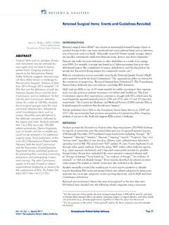
7 Moreover, x-rays have been The following PA-PSRS reports indicate misinter-pretations by nonradiologists: A house physician in-serted a Keofeed tube in a geriatric patient. Both the nurse and physi-cian confirmed placement by auscultating insuf-flated air. The physician confirmed placement af-ter reading the x-ray. Tube feedings were begun. The patient was found dead. A Dobhoff tube was placed by a house physi-cian. The x-ray was read and placement confirmed. Tube feedings were initi-ated. The patient experi-enced respiratory dis-tress. A review of the x-ray showed that the Feeding tube was in the main bronchus. Confirmation that the Feeding tube is properly placed in the stomach or small bowel involves documenting the following on a chest x-ray: 1. The tube follows a straight course down the midline of the chest to a point below the diaphragm.
8 2. The tip of the tube is below the diaphragm. Figure 1. Chest Radiograph Representing Properly Placed nasogastric Feeding Tube with Tip Visible The tube follows a straight course down the midline of the chest to a point below the diaphragm. The tube does not follow the path of a bronchus. Tube is not coiled anywhere in the chest. The tip of the tube is below the diaphragm. Confirming Feeding Tube placement : Old Habits Die Hard (Continued) Page 3 Reprinted from the PA-PSRS Patient Safety Advisory Vol. 3, No. 4 (Dec. 2006) 2006 Pennsylvania Patient Safety Authority 3. The tube is not coiled anywhere in the chest. 4. The tube does not follow the path of a If the tube is intended to be placed in the small bowel, an abdominal x-ray is needed to determine where the ports are situated. Small bowel feedings are needed when patients cannot tolerate gastric feedings because of significantly delayed gastric emptying, demonstrated chronic aspiration of gastric contents, or a known incompetent lower esophageal sphincter.
9 In the United Kingdom, the National Patient Safety Agency does not recommend the routine use of x-ray for nasogastric tube placement confirmation, reserving it for patients at high risk for misplacement of the nasogastric tube, such as the critically ill or Endoscopy and Fluoroscopy Both endoscopy and fluoroscopy accurately verify placement of Feeding tubes , but these methods can be cost-prohibitive, time-consuming, and pose addi-tional risks, such as transporting patients to special procedures areas or imaging departments. Because fluoroscopy produces clinically significant radiation exposure, this technique is used for Feeding tube placement only as a last pH Testing Another reliable method for ongoing tube placement verification is determining the pH of the fluid aspi-rated from Feeding tubes . Gastric fluid is usually acidic, with a pH less than or equal to Respira-tory secretions are almost always alkaline, with a pH greater than or equal to 6.
10 In a large study of 1,284 aspirates from Feeding tubes , all samples from the lungs had a pH greater than or equal to If the pH of the Feeding tube aspirate is greater than or equal to 6, the tube may be inadvertently located in the respiratory ,17 However, several conditions can affect the pH of aspirates, resulting in misinterpretation of the place-ment of a Feeding For example, respiratory secretions may be acidic in patients with esophag-eal rupture, acid reflux, or a pleural infection such as Feeding tube aspirates are usually alkaline if the tube is in the small bowel or the patient is Also, gastric pH will rise temporarily when the patient is receiving acid-inhibiting medications ( , histamine2-antagonist, proton pump inhibitor) or when tube feedings are in Confirming Feeding Tube placement : Old Habits Die Hard (Continued) Figure 2.