Transcription of Criteria for Mammographic Image Assessment
1 8/31/20161 Criteria for Mammographic Image Assessment Faculty:Deborah Thames, RT (R) (M)Mammography SpecialistHouston, TexasCourse Topics:Review of Positioning Criteria for Routine MammogramsMedio lateral Oblique (MLO) Assessment of the MLO Image by means of: Posterior nipple lineInframammary foldClinical Image evaluation using MLO positioning criteriaEvaluation of adequate compression on MLO imagesCranio-caudal (CC) Assessment of CC Image by means of:pectoral muscleposterior medial breastClinical Image evaluation using CC positioning criteriaEvaluation of adequate compression on CC imagesACR Criteria for Mammographic Image Assessment ACR Criteria for Mammographic Image Assessment Course Topics:Review of Technical Aspects with Clinical Image ExamplesPositioning* Compression* Exposure level Contrast Sharpness Noise Artifacts Exam identificationAdministrative ConcernsLabeling 8/31/20162 National Statistics (MQSA)8/31/20163 Mammographic images submitted for accreditation review must be.
2 Negative (BI-RADS Assessment Category 1) No benign (Category 2) No incomplete (Category 0) If the facility only performs diagnostic exams and cannot submit negative images, they should call the ACR for assistance Cases must be examples of the facility s best work Images must be from actual patients and must have been formally interpreted Images from models or volunteers are not acceptable Mammographic Clinical Image Criteria for Accreditation Submissions8/31/20164 Criteria for Mammographic images submitted for accreditation review: Complete breast must be imaged in a single exposure on each projection , any breast tissue missing is considered an automatic failure. digital images must be as close to true size as , with no minification or magnification Both screen - film and digital images must be labeled with the MQSA-required identification information Lead interpreting physician must review and approve the clinical images submitted Electronically submitted images must be processed marked For Presentation Mammographic Clinical Image Criteria for Accreditation SubmissionsClinical Images & Image Quality Interpreting PhysiciansPhysicians interpreting mammograms for the facility shall follow the facility procedures for corrective action when the images they are asked to interpret are of poor quality.
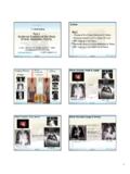
3 There should be a procedure in place to follow when images do not meet the established clinical standards Clinical Image ParametersPercentages are in order of resultant causes of clinical Image failure Positioning 20% Exposure 15% Compression 14% Sharpness 13% Contrast 13% Artifacts 11% Labeling ID 8% Noise 5% 8/31/20165 Most Common Positioning Errors Poor visualization of posterior tissue Sagging breast Inadequate amount of pectoralis major muscle on Image Excessive exaggeration on the craniocaudalview Skin foldsCranial Caudal View Pectoralis muscle is visualized in only 30-40 % of patients according to ACR manual, but with new positioning skills, more like 60 percent.
4 When the muscle is not included, the measurement of the PNL should be done Medial vs lateral tissue Nipple in profile, good to have nipple in profile on all views for ACR Look for variation in nipple location, must be Oblique View Pectoralis muscle included to the PNL Muscle should be wide and convex Inframammary Fold (IMF) seen on Image Retroglandular fat included Look for variation in nipple location8/31/20166 Sagging breast, cut off bottom of breast on film for RT MLO viewPositioning Other body parts projected over breast Nonstandard angulation MLO 30-60 degree Posterior nipple line on craniocaudal view not within 1 cm of that on the mediolateral oblique view Breast positioned too high on Image receptor8/31 CC measured Lt CC measured mm8/31/20169 Inadequate amount of pectoralis major muscle on Image Skin fold and too much exaggeration on RtCC view Skin fold in RT MLO8/31/201610 Nipple not in profile Other body parts projected over breast area8/31/201611 Breast too high on Image receptor8/31/201612 CompressionResults of inadequate compression Poor separation of parenchymal densities Non-uniform
5 Exposure levels Patient motion Poor separation of parenchymal densities Patient Motion8/31/2016138/31/201614 Contrast Inadequate contrast Excessive contrast Contrast Image shall permit differentiation of subtle tissue density differences Must watch Window Leveling and width, especially if you don t you don t have a post processing algorithm GE has premium view and fine Visually striking mottle pattern Noise-limited visualization of detail Noise in the Image shall not obscure breast structures or suggest the appearance of structures not actually Punctate or lint with film screen Scratches or pickoff with film screen Roller marks with film screen Grid-related artifacts film screen / digital Hair, deodorant film screen / digital Image handling film screen Image fogging film screen Poor screen - film alignment film screen Dead pixels artifacts digital Lag and/or ghosting digital Streaking and misread columns digital Grid related artifacts Hair8/31/201616 Deodorant Image fogging8/31/201617 Skin FoldsSome FFDM Artifacts8/31/2016188/31/2016198/31/2016 208/31/201621 Exposure Generalized underexposure Generalized overexposure Inadequate penetration of dense areas Excessive penetration of radiolucent areas Exposure level shall be adequate to visualize breast structures.
6 Underexposed Image Overexposed image8/31/201622 Inadequate penetration of dense areaCompression Compression shall be applied in a manner that minimizes the potential obscuring effect of overlying breast tissue and motion artifact8/31/201623 Sharpness Poor delineation of linear structures Poor delineation of feature margins Poor delineation of microcalcifications Margins of normal breast structures shall be distinct and not blurred Delineation of linear structures8/31/201624 Delineation of calcificationsLabeling of Mammograms Mammography films are medical documents. To make sure no misinterpretation of films,label films information on labeling are required by federal law and some information is recommended. Required by Federal LawOn Name Label MQSA-Required Mammographic Image Identification 1.
7 Name of patient (first and last) 2. Additional patient identifier ( , medical record number or social security number; date of birth is less desirable) 3. Date of examination 4. Standardized view and laterality codes placed on the Image in a position near the axilla 5. Facility name and location (must include city, state, and zip code) 6.
8 Technologist identification 7. Cassette/ screen identification 8. Mammography unit identification, if more than one unit in the facility 8/31/201625 Required by Federal Law Radiopaque Markers indicating laterality like Left or Right on film screen / digital must have lt/rt Projection view as in CC or MLO, The Technologist who performed the examinations, may be technologist initials or a technologist by Federal Law Cassette/ screen identification to identify screens even with CR Mammography. Mammography unit number if there is more than one unit in the recommended A flash card patient ID system for more permanent measures.
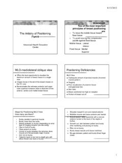
9 Flash system is not acceptable if any information is illegible, does not fit, or is lobsided, causing cut-off of information. 8/31/201626 Recommended Separate Date stickers as they allow the for the date to be easily read. Technical factors Target filters KvP mAs Exposure Time Compression force Compressed breast thickness Degree of obliquity Remember Full Field digital Units can submit their ACR film Accreditations in Hard Copy Format or electronically8/31/201627 Failure verses Deficiency* A first deficiency is not a does not notify the do not have to stop doing mammograms in your corrective actions on your own.* Repeat deficient test less than 2 months MQSA* Reinstate if more than 2 months on MQSA cert. * Appeal* WithdrawSum it up8/31/201628 Good mammogramBad mammography8/31/201629 Good mammogramBad mammographyGood mammogram8/31/2016308/31/2016318/31/2016 32Tw o C a t e g o r i e sA/B category oneC/D category two8/31/201633 Examples of ACR Images Remember all has to be the very best images from your facility8/31/201634 Checks on CC Posterior tissue/fat and possible muscle Nipple perfectly centered Nipple in profile No folds Good compression at least 20lbs or more Separation of densities Good ContrastChecklist on MLO Tail of breast on Image Nipple in profile Retro glandular Fat from Clavicle to 6thrib where IMF PNL line within 1 CM of CC IMF must be on Image Densities are well separated Muscle is wide
10 Superiorly with a convex border. Center of Image should be 2 cm above nipple Good contrast8/31/201635 FULL RESULT:CLINICAL INDICATION:Patient is a 52 year old female and is seen for digital SCREENING MAMMOGRAMD igital Screening Mammogram evaluated with Computer Aided Detection (CAD).COMPARISON:The present examination has been compared to prior imaging studies performed atCancer Center on 03/12/2010, 07/15/2011 and 04/12 : The breasts are heterogeneously dense. This may lower the sensitivityof views are recommended to include more posterior tissue in the :Findings in both breasts require additional evaluation. The following views willneed to be repeated for technical reasons; (bilateral craniocaudal).BI-RADS Category 0:Additional Imaging Evaluatio