Transcription of DETERMINATION OF DNA CONCENTRATION AND PURITY ¶ ¶ …
1 BIOTECHNOLOGY I DNA CONCENTRATION AND PURITY Eilene Lyons Revised 1/12/2010 Page 2-1 LAB 2 DETERMINATION OF DNA CONCENTRATION AND PURITY STUDENT GUIDE GOAL The goal of this lab is to give experience in one technique for extracting chromosomal DNA from plant tissue and in DETERMINATION of chromosomal DNA quality and quantity. OBJECTIVES After completion, the student should be able to: 1. List and explain the steps of chromosomal DNA isolation. 2. Determine the quality and PURITY of chromosomal DNA using gel electrophoresis and UV spectrophotometry. 3. Calculate DNA CONCENTRATION and from agarose gel electrophoresis results 4. Calculate DNA CONCENTRATION from UV absorbance results. 5. Use laboratory protocol reference books and the Internet to locate information for use in the laboratory. TIMELINE This lab will take 1 laboratory period to: 1. Determine DNA quality, CONCENTRATION and PURITY using agarose gel electrophoresis 2. Determine DNA CONCENTRATION and PURITY using UV absorbance BACKGROUND Nucleotide Structure The building block or monomer of nucleic acid is the nucleotide.
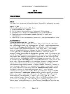
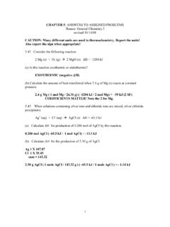
2 Nucleotides consist of three parts: (1) a five-carbon sugar, (2) a nitrogenous base, and (3) a phosphate group. There are two kinds of nucleic acids: DNA and RNA. The monomers of DNA and RNA vary slightly. RNA contains the five-carbon sugar ribose; DNA contains the five carbon sugar deoxyribose, which has one less oxygen. See Figure 1, below and nucleotide models in the lab. Note that the carbons in the sugar molecules are designated with numbers 1 -5 (pronounced 1 prime to 5 prime) to distinguish them from the carbons in the nitrogenous bases. Also, the 5 carbon in the sugar is not part of the ring structure, but sticks out above it. 0- HO P O CH2 0O OH HHC NCHCNH2 N COSUGARBASEPHOSPHATE5 4 1 2 3 A 0- HO P O CH2 0O OH OHSUGARBASE (URACIL)5 4 1 2 3 N C NHCOCH HCOB Figure 1.
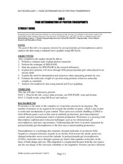
3 A. DNA Nucleotide; B. RNA Nucleotide BIOTECHNOLOGY I DNA CONCENTRATION AND PURITY Eilene Lyons Revised 1/12/2010 Page 2-2 The nitrogenous bases of DNA include adenine (A), thymine (T), guanine (G), and cytosine (C). Cytosine and thymine are single-ringed structures called pyrimidines; adenine and guanine are double- ringed structures called purines (see Figure 2). RNA contains the same nitrogenous bases as DNA except for thymine. Uracil replaces thymine in RNA. Uracil, like thymine, is a single-ringed molecule. These nitrogenous bases are able to hydrogen bond with their complement, another base that forms the same number of hydrogen bonds. A purine will always hydrogen bond with a pyrimidine. Cytosine and Guanine are complementary, forming three hydrogen bonds between them. Adenine and thymine (uracil in RNA) are complementary, forming two hydrogen bonds between them. This hydrogen bonding between complementary bases is referred to as base pairing.
4 One set of nucleotides hydrogen bonded together at their complementary bases is referred to as a base pair. See Figure 2 and models in the lab. DNA Structure and Function DNA is a double stranded molecule made up of two polymers of nucleotides. The nucleotides of each polymer are bonded together at the sugar of one nucleotide and the phosphate of the next. The two polymers are joined in the center by hydrogen bonds between the nitrogenous bases. DNA has the shape of a ladder, with the sides of the ladder composed of alternating phosphates and sugars while the rungs of the ladder are the hydrogen-bonded bases. The ladder is twisted so that it resembles a spiral staircase and therefore, is called a double helix. DNA carries the master blueprint for heredity in the form of a code. Within a gene that codes for a protein, sets of three nucleotides code for a particular amino acid.
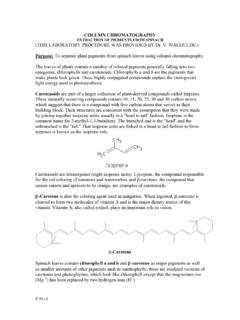
5 The 64 different three-nucleotide combinations of the four nucleotides of DNA (A,T,G,C) are called the genetic code. The sequence of 3-nucleotide codons in the DNA serves as the blueprint for the primary structure (the amino acid sequence) of a protein. O5 4 1 2 3 PSGO5 4 1 2 3 PSCO5 4 1 2 3 PSTO5 4 1 2 3 PSA Figure 2. Base Pairing of Complementary Nucleotides BIOTECHNOLOGY I DNA CONCENTRATION AND PURITY Eilene Lyons Revised 1/12/2010 Page 2-3 Chromosomal DNA Extraction The technique for chromosomal DNA extraction depends on the type of cell or tissue being used. Since plant cells have a fibrous cell wall consisting of cellulose and pectin, plant tissue must be ground, macerated and sometimes heated to 65C to rupture the cells releasing the DNA. With animal and gram positive bacterial cells, enzymes such as Protease K, lysozyme or papain (a protein-cleaving enzyme derived from papaya and certain other plants) are used.
6 More extreme measures must be taken to remove the thick peptidoglycan layer from gram positive bacteria. One technique is to vortex the cells with glass beads and a detergent such as sodium dodecyl sulfate (SDS). Gram negative cells, such as E. coli, can be lysed using SDS without vortex mixing. The SDS dissolves the phospholipid bilayer of cell membranes and also the nuclear membrane in eukaryotic cells. To precipitate the DNA, a salt such as ammonium, sodium or potassium acetate is added and then alcohol is added (ethanol or isopropanol, depending upon the protocol). The salt must be added first because DNA will not precipitate in alcohol otherwise. Since cold enhances the precipitation of DNA, -20 C alcohol can be used and the DNA/alcohol mixture can be incubated for a short time (5-20 minutes) at -20 C. Pure DNA is clear and nearly transparent, but in many extractions, it appears as a cloudy mucous-like mass of strands floating in the alcohol.
7 Protein gives DNA this milky, opaque appearance. With plant DNA extraction (especially from fruit), there can also be polysaccharide contamination, visible as a clear jelly-like mass. Both protein and polysaccharide contamination must be removed because each inhibits the action of enzymes, the molecular tools of biotechnology. If floating white flakes appear in the alcohol, the DNA was sheered into small pieces and is not usable for further genetic manipulation. The DNA precipitate is then centrifuged to separate it from the alcohol. The DNA pellet is resuspended in Tris buffer containing Ethylenediamine tetraacetic acid (EDTA). The EDTA chelates (binds) divalent cations that are cofactors necessary for Dnases, the enzymes that degrade DNA. DNA resuspended in Tris-EDTA should be stored short term at -20 C or long term at -80 C. Chromosomal DNA Cleanup Good quality DNA is not only intact but is also clean free of salt, proteins and polysaccharides that inhibit DNA-modifying enzymes.
8 To remove proteins, an affinity gel can be employed to either bind the DNA while the protein is washed away or bind the protein while the DNA is washed off and collected. Equilibrated phenol (saturated with 10 mM Tris-HCl, pH and 1 mM EDTA to increase the phenol s pH to , which is required for DNA extractions), chloroform or a combination of these two organic solvents, are used to wash the DNA. The non-polar portions of the proteins are soluble in organic solvents so they are dragged down and out of the aqueous DNA solution as the dense solvents sink to the bottom of the tube. The hydrophilic parts of the proteins get dragged down and out of the aqueous solution and end up in a whitish layer, or interface, floating on top of the organic phase at the bottom. A small amount of isoamyl alcohol can be used to separate the two phases more efficiently, making it easier to draw off the aqueous DNA layer at the top. Phase Lock Gel can also be used to separate the aqueous phase for easy removal.
9 This is a proprietary product that eliminates protein contamination by migrating during centrifugation to form a tight seal trapping the interface material and organic phase below the aqueous phase. The aqueous phase is then simply decanted or pipetted off the top and transferred to another tube (Murphy and Hellwig). BIOTECHNOLOGY I DNA CONCENTRATION AND PURITY Eilene Lyons Revised 1/12/2010 Page 2-4 Polysaccharides are precipitated with ethanol at a CONCENTRATION lower ( volumes) than that used to precipitate DNA (2 volumes). The salt CONCENTRATION of the aqueous phase is critical here no more than M or the excess salt will cause the polysaccharides to remain in solution (Michaels, John, Amasino). Any remaining salt is removed by washing the pellet of DNA with isopropanol or ethanol. RNA will also be present, but does not interfere with the action of DNA-modifying enzymes used in genetic engineering. RNA can give an inflated DNA CONCENTRATION , so it is often removed by incubation with RNase, an enzyme that degrades RNA, prior to taking UV absorbance readings.
10 There are commercial DNA clean-up kits available (Qiagen, Zymo Research, Sigma-Aldrich) that work well to remove contamination. DETERMINATION of DNA Quality and Quantity After the extraction procedure, the DNA is checked to verify that it is intact and clean of cellular contaminants. DETERMINATION of the quality and CONCENTRATION can be accomplished in two ways: gel electrophoresis and UV spectrophotometry. Gel Electrophoresis DNA can be diluted and run on an agarose gel. By running a molecular weight marker of known CONCENTRATION , the extracted DNA CONCENTRATION can be determined. The appearance of the DNA on the gel can also reveal if it is clean and intact. Agarose is a derivative of agar, a polysaccharide derived from algae. DNA fragments, including molecular weight markers, are often heated to 65C prior to electrophoresis to straighten any loops formed along the length of the molecules so that migration through the gel is uniform.