Transcription of EDUCATIONAL COMMENTARY BASIC – VAGINAL …
1 EDUCATIONAL COMMENTARY BASIC VAGINAL WET PREP EDUCATIONAL COMMENTARY is provided through our affiliation with the American Society for Clinical Pathology (ASCP). To obtain FREE CME/CMLE credits click on Earn CE Credits under Continuing Education on the left side of the screen. **Florida licensees, please note: This exercise will appear in CE Broker under the specialty of hematology. Learning Outcomes On completion of this exercise, the participant should be able to describe characteristics used to identify Trichomonas vaginalis in a VAGINAL wet prep; describe characteristics used to determine whether an epithelial cell is a clue cell; describe how to differentiate small yeast cells from red blood cells; and list the advantages and disadvantages of the VAGINAL wet prep.
2 BASIC VAGINAL Wet Prep In an age of increasingly complex laboratory testing, certain low-tech laboratory tests remain valuable to the clinician. The BASIC VAGINAL wet preparation or VAGINAL wet prep used to check for the presence of Trichomonas vaginalis, yeasts, and clue cells is one such test. In the 17th and 18th centuries when Antonie van Leeuwenhoek and Robert Hooke began to explore the microscopic world with the first microscopes, they already had the tools to perform VAGINAL wet preps. The equipment required consists of a light microscope, glass slides, coverslips, and saline. This test can be done in the physician office laboratory as well as in the most modern hospitals.
3 It can be performed in a timely manner and with minimal equipment. Although the BASIC VAGINAL wet prep can performed quickly with readily available materials, it requires personnel trained in microscopy. The VAGINAL wet prep also has the disadvantage of lacking sensitivity and specificity. It is not useful in diagnosing vaginitis caused by some pathogens such as herpes simplex virus. Other tests that are valuable when done in conjunction with the VAGINAL wet prep include the pH of the discharge and the whiff test. The appearance of the discharge is also helpful in determining the diagnosis. Vaginitis or vulvovaginitis may be caused by a variety of microbial organisms.
4 The most common sources of vaginitis include yeast (predominantly Candida albicans), Gardnerella vaginalis, and the parasite Trichomonas vaginalis, all of which can be observed using BASIC light microscopy. Bacterial vaginosis American Proficiency Institute 2015 2nd T est Event EDUCATIONAL COMMENTARY BASIC VAGINAL WET PREP (cont.) caused by G vaginalis is diagnosed by the presence of clue cells, VAGINAL candidiasis by identifying budding cells or long pseudohyphae, and trichomoniasis by observing motile T vaginalis trophozoites. VAGINAL Wet Prep Procedure Materials required include cotton or synthetic-tipped swabs, to 1 mL of saline, microscope slides with coverslips, and a light microscope with 10X and 40X objectives.
5 VAGINAL discharge is collected on a swab and placed into a test tube containing between and 1 mL of saline. The sample is not stained or mixed with any reagent. The sample should be examined within 1 hour of collection. One drop is placed onto a glass microscope slide and covered with a coverslip. The slide is examined on low (10X) power under low light. The 40X objective may be used to confirm the presence or absence of T vaginalis, G vaginalis, and yeasts. An alternative technique is to add a drop of saline to a slide and add a small amount of discharge. The slide is covered with a coverslip and examined using the 10X and 40X objectives.
6 It may be helpful to gently warm the sample to increase the motility of T vaginalis. Trichomonas vaginalis Trichomonas vaginalis is a single-cell, flagellated parasite that replicates by binary fusion. It has no known cyst stage. It resides in the female lower genital tract and in males in the urethra and prostate, and is spread by sexual intercourse. Its only known host is humans. T vaginalis is identified by detecting motile flagellates. T vaginalis is slightly larger than a white blood cell and should have flagellar (axostyle and undulating membrane) motility. If motility is present, the sample may be reported as positive for the presence of T vaginalis.
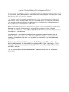
7 It is important to examine the sample as soon after collection as possible, as the organism will lose its motility with delay. It has been determined that motility is present 100% of the time at 30 minutes, 99% at 1 hour, and decreases 3% to 15% for each hour thereafter. The sensitivity of the test also depends on the amount of organisms in the sample. When the organism is no longer motile, it rounds up, internalizing the flagella and becoming indistinguishable from a white blood cell (Figure 1). This organism is highly susceptible to drying, so a sample received on a slide without saline should be rejected.
8 Because the trophozoite of Trichomonas hominis resembles that of T vaginalis, any sample contaminated with stool should also be rejected. Trichomonas vaginalis causes a vaginitis that is characterized by a purulent VAGINAL discharge in women. It is often asymptomatic in men. The sequelae of T vaginalis infection may include complications in pregnancy, cervical cancer, and a predisposition to infection with human immunodeficiency virus (HIV). Treatment often consists of metronidazole or tinidazole. American Proficiency Institute 2015 2nd T est Event EDUCATIONAL COMMENTARY BASIC VAGINAL WET PREP (cont.)
9 Other diagnostic testing for T vaginalis includes culture, molecular methods, direct fluorescent antigen (DFA) tests, and antigen-detection tests. Culture methods are highly sensitive, but it can take up to a week for results to be available. Molecular tests are also highly sensitive and specific but are usually expensive and require specialized equipment. Direct fluorescent antigen testing also requires specialized equipment and specially trained personnel. Along with the VAGINAL wet prep, antigen-detection tests are better suited for physician office laboratories. For the detection of T vaginalis, the VAGINAL wet prep has a sensitivity of approximately 60% and specificity of 100%.
10 Figure 1. Trichomonas vaginalis. The characteristic twisting, rotating appearance is highly specific for T vaginalis. It is important to keep the time from collection to testing as short as possible: ideally, within 1 hour of collection. As the sample ages, the motility of the organism decreases. Once the Trichomonas organism is no longer motile, it cannot be distinguished from a white blood cell. Gardnerella vaginalis Gardnerella vaginalis is an anaerobic, gram-positive coccobacillus. Because of its thin cell walls, G vaginalis often stains as gram-variable. It may be present at low levels in normal VAGINAL flora.