Transcription of Electroencephalography - Rice University
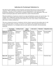
1 Electroencephalography1 ElectroencephalographyAn EEG recording net (Electrical Geodesics,Inc.[1] ) being used on a participant in a brainwave spike and wave discharges monitoredwith (EEG) is the recording of electrical activityalong the scalp produced by the firing of neurons within the brain.[2] Inclinical contexts, EEG refers to the recording of the brain'sspontaneous electrical activity over a short period of time, usually20 40 minutes, as recorded from multiple electrodes placed on thescalp.
2 In neurology, the main diagnostic application of EEG is in thecase of epilepsy , as epileptic activity can create clear abnormalities ona standard EEG study.[3] A secondary clinical use of EEG is in thediagnosis of coma, encephalopathies, and brain death. EEG used to bea first-line method for the diagnosis of tumors, stroke and other focalbrain disorders, but this use has decreased with the advent ofanatomical imaging techniques such as MRI and of the EEG technique include evoked potentials (EP),which involves averaging the EEG activity time-locked to thepresentation of a stimulus of some sort (visual, somatosensory, orauditory).
3 Event-related potentials refer to averaged EEG responsesthat are time-locked to more complex processing of stimuli; thistechnique is used in cognitive science, cognitive psychology, andpsychophysiological of EEG activityThe electrical activity of the brain can be described in spatial scalesfrom the currents within a single dendritic spine to the relatively grosspotentials that the EEG records from the scalp, much the same way thateconomics can be studied from the level of a single individual'spersonal finances to the macro-economics of nations.
4 Neurons, ornerve cells, are electrically active cells that are primarily responsiblefor carrying out the brain's functions. Neurons create action potentials,which are discrete electrical signals that travel down axons and causethe release of chemical neurotransmitters at the synapse, which is anarea of near contact between two neurons. This neurotransmitter thenactivates a receptor in the dendrite or body of the neuron that is on theother side of the synapse, the post-synaptic neuron.
5 Theneurotransmitter, when combined with the receptor, typically causes anelectric current within the dendrite or body of the post-synaptic neuron. Thousands of post-synaptic currents from asingle neuron's dendrites and body then sum up to cause the neuron to generate an action potential. This neuron thensynapses on other neurons, and so reflects correlated synaptic activity caused by post-synaptic potentials of cortical neurons. The ionic currentsinvolved in the generation of fast action potentials may not contribute greatly to the averaged field potentialsrepresenting the EEG.
6 [4] [5] More specifically, the scalp electrical potentials that produce EEG are generally thoughtto be caused by the extracellular ionic currents caused by dendritic electrical activity, whereas the fields producingmagnetoencephalographic signals[6] are associated with intracellular ionic currents .[7]Electroencephalography2 The electric potentials generated by single neurons are far too small to be picked by EEG or MEG.[5] EEG activitytherefore always reflects the summation of the synchronous activity of thousands or millions of neurons that havesimilar spatial orientation.
7 Because voltage fields fall off with the square of the distance, activity from deep sourcesis more difficult to detect than currents near the skull.[8]Scalp EEG activity shows oscillations at a variety of frequencies. Several of these oscillations have characteristicfrequency ranges, spatial distributions and are associated with different states of brain functioning ( , waking andthe various sleep stages). These oscillations represent synchronized activity over a network of neurons. The neuronalnetworks underlying some of these oscillations are understood ( , the thalamocortical resonance underlying sleepspindles), while many others are not ( , the system that generates the posterior basic rhythm).
8 Research thatmeasures both EEG and neuron spiking finds the relationship between the two is complex with the power of surfaceEEG only in two bands that of gamma and delta relating to neuron spike activity.[9]Clinical useA routine clinical EEG recording typically lasts 20 30 minutes (plus preparation time) and usually involvesrecording from scalp electrodes. Routine EEG is typically used in the following clinical circumstances: to distinguish epileptic seizures from other types of spells, such as psychogenic non-epileptic seizures, syncope(fainting), sub-cortical movement disorders and migraine variants.
9 To differentiate "organic" encephalopathy or delirium from primary psychiatric syndromes such as catatonia to serve as an adjunct test of brain death to prognosticate, in certain instances, in patients with coma to determine whether to wean anti-epileptic medicationsAt times, a routine EEG is not sufficient, particularly when it is necessary to record a patient while he/she is having aseizure. In this case, the patient may be admitted to the hospital for days or even weeks, while EEG is constantlybeing recorded (along with time-synchronized video and audio recording).
10 A recording of an actual seizure ( , anictal recording, rather than an inter-ictal recording of a possibly epileptic patient at some period between seizures)can give significantly better information about whether or not a spell is an epileptic seizure and the focus in the brainfrom which the seizure activity monitoring is typically done: to distinguish epileptic seizures from other types of spells, such as psychogenic non-epileptic seizures, syncope(fainting), sub-cortical movement disorders and migraine variants.


![How to Interpret and EEG and Its report.ppt [Read-Only]](/cache/preview/9/a/8/4/5/3/5/0/thumb-9a84535099038c890ee91bcb8802c130.jpg)