Transcription of ENVIRONMENTAL CONDITIONS & BACTERIA GROWTH
1 ENVIRONMENTAL CONDITIONS &. BACTERIA GROWTH . Many ENVIRONMENTAL CONDITIONS can affect microbial GROWTH ---temperature, pH, osmotic pressure, radiation, and barometric pressure. PH is the value determined by the concentration of hydrogen ions in a liquid, represented by the formula: [H+] = moles of H+/liter The value represents a base 10 logarithm, and pH therefore defines a logarithmic scale of acidity. The difference in numbers on the pH scale (0 to 14) is 10-fold. That is, pH 8 is 10X. more basic than pH 7 and pH 3 is 100X more acidic than pH 5. Water naturally dissociates into H+ and OH ions with equal concentrations of 1 10 7 M, hence a pH of 7 is neutral.
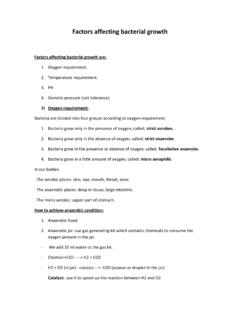
2 Less than 7 is acidic (the more extreme the number, the greater the H ion), while higher than 7 is basic (the more extreme, the less the H ion). Most BACTERIA live within the pH range of 4-9. Acidophiles and alkalinophiles are the extreme microorganism groups. Fungi often can live at lower pH CONDITIONS , citrus fruit decay. Like your human cells, most BACTERIA optimally grow in isotonic CONDITIONS . However, many microbes, particularly fungi, can tolerate hypertonic CONDITIONS produced by high salt or high sugar concentrations. The flow of water is controlled by the osmotic pressure of the environment, and that is determined by the concentration of solute molecules.
3 Osmophilic microorganisms tend to be fungi. Those organisms that require high salt are called halophilic, some requiring concentrations of 15-20% NaCl. Many BACTERIA can tolerate high salt concentrations (halotolerant), although they do not really require the salt to grow. Hypertonic ENVIRONMENTAL CONDITIONS cause the cells to dehydrate as water osmoses out of the cell. In the following exercise, you will look at the effect of various salt concentrations on bacterial GROWTH . Generally, the degree of inhibition of GROWTH will depend on the type of dissolved particles (salt, sugar, etc.), its concentration, and the type of microorganism.
4 Radiation is often used to control microbial GROWTH (rooms, foods, packaged products, etc.). The killing ability of radiation is due to mutations in the DNA produced by the radiation. Two major forms of electromagnetic radiation can be mutagenic ionizing radiation (X-rays, gamma) and ultraviolet. Ionizing is the more potent since it is much shorter wavelength, causing electrons to be pulled off of DNA molecules and oxidizing it. Ultraviolet radiation exerts it effect by causing adjacent pyrimidine nitrogenous bases (commonly thymine bases). to bond with each other, meaning that the strand cannot effectively attach to its complementary strand.
5 These thymine dimers affect the replication of the DNA in the dividing cell. There are different kinds of ultraviolet, the most germicidal being UV-C (because it penetrates the best). Ionizing radiation has much greater kill Fall 2011 Jackie Reynolds, Richland College, BIOL 2420. ability because of its penetration (short wavelength). Ultraviolet radiation does not generally penetrate clothes, glass, or plastic. Obviously you will have to remove the tops of the plastic petri dishes when exposing the BACTERIA to the UV. Cells have repair mechanisms that counteract these mutations, but if the mutagenic agent is producing the mutations faster than can be fixed, the cell will not live.
6 The cell repairs the mutation in different ways: 1. excising the incorrect section of the DNA with enzymes 2. producing an enzyme that uses energy of visible light to split the thymine dimmers The latter method is called photoreactivation. OBJECTIVES: Identify the effects of ultraviolet radiation on bacterial GROWTH . Identify the effects of pH on bacterial GROWTH . Identify the effects of osmotic pressure on bacterial GROWTH . MATERIALS NEEDED: per table OSMOTIC PRESSURE exercise 1 TSA plate each of (regular TSA), 5%, and 10% NaCl concentrations PH exercise 1 TSB pH 3, 7, and 10 for each organism UV radiation exercise 4 X 6 index cards 5 TSA plates 8 sterile cotton swabs UV lamp (bulb should be positioned 18 inches above the table top).
7 TSB cultures (same density of cells in each) of various BACTERIA (your instructor will assign). a spore suspension of Bacillus subtilus (for UV study only). THE PROCEDURES: OSMOTIC PRESSURE (each table uses all 3 organisms) different inoculation patterns 1. Each table will obtain one TSA plate of each salt concentration and divide the plate into 3 sections (3 BACTERIA in 3 sections). 2. Inoculate a section of each agar plate with a loopful of your 3. organisms, trying to use the same amount of inoculum on each plate and inoculated the same way for consistency. 3. Incubate the plates in incubator recommended by instructor. ULTRAVIOLET RADIATION (each table will use1 organism).
8 1. Label your 5 plates with the name of the bacterium and exposure time (remember that 1 plate, exposed for 60 seconds, will have a lid during UV exposure be sure to label appropriately). 2. 2. Dip the swab in your assigned culture. Use the swab to smear the culture all over the ENTIRE plate of agar. Streak back and forth in a DENSE zig-zag pattern repeatedly to cover every bit of agar. 3. Take your plates to the UV lamp and place on the table top (18 inches below the bulb). 4. Do NOT remove the tops of the petri dishes until READY to expose to UV. You should be able to get all of the plates directly under the lamp. 5. Marking the time on your watch or a clock as time ZERO), remove the petri dish cover and expose the plate to the UV for the assigned amount of time.
9 Do the same with all of the other agar plates, except the 10 minute exposure (see directions below). exposure times for plates: 10 seconds 60 seconds (2 plates 1 exposed, 1 covered with lid). 5 minutes 10 minutes (Place the index card over of the agar plate at a right angle to the swab lines, on top of the petri dish with cover on it, Remove the plate cover when ready to start timing). WARNING: Do not look directly into the UV light as it can cause damage to your eyes. Do not expose your direct skin to the UV. 6. Remove the each plate from the UV and cover the plate with the Petri dish lid. 7. Incubate all plates at 25C. PH (each table will use 1 organism).
10 1. Each table will obtain 3 TSB tubes of pH 3, 7, and 10. Your table will be assigned ONLY 1 bacterium to inoculate all of the pH broths. 2. Inoculate each TSB with a loopful of your organism. INTERPRETATION: OSMOTIC PRESSURE agar plates: During the next lab period you will determine the effect of salt on the GROWTH of your bacterium. Quantify the amount of GROWTH by comparing the density of the organisms on the plates. RECORD DATA IN TABLE BELOW, including other tables' data. - no GROWTH +1 very light GROWTH +2 medium GROWTH +3 heavy GROWTH 3. UV RADIATION agar plates Compare the 60 second plate that had a lid during UV exposure with the one that was exposed.