Transcription of Feline ophthalmology Part 2: clinical presentation …
1 Continuing education Volume 59 (4) : april, 2006 irish Veterinary Journal 223 Feline ophthalmology part 2: clinical presentation and aetiology of common ocular conditionsnatasha MitchellDownland Veterinary Group, 71 Havant Rd., Emsworth, Hampshire, PO9 7NZ, EnglandTel: +44 1243 377 141; Email: this, the second par t of our series on Feline ophthalmology , emphasis is placed on the clinical appearance of the common conditions which regularly present in practice. Performing the thorough examination outlined in part 1 will detect abnormalities. Using the information in Par t 2 should help the clinician to correctly identify a condition, allowing treatment and prognosis to be condition is manifested as congestion or oedema (chemosis) of the conjunctiva.
2 It may be accompanied by an ocular discharge. Secondary conjunctivitis may occur with other ocular conditions, such as uveitis and glaucoma. In cats, conjunctivitis is frequently caused by an infectious agent, and multiple agents may be involved. Resident ocular bacterial flora is present; therefore, a bacteriology swab does not always produce a result with a causative organism. Staphylococcus aureus, Staphylococcus epidermidis, Streptococcus spp and Corynebacterium spp are commonly isolated commensal organisms from the normal Feline conjunctiva and eyelids (Gerding and Kakoma, 1990).Many upper respiratory tract pathogens cause conjunctivitis.
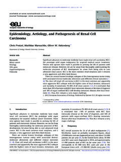
3 Feline herpesvirus-1 (FHV-1) can infect domestic and wild cats (Maggs et al.,1999a,b). The virus is considered to be responsible for nearly half of all upper respiratory tract infections in these species. Replication occurs in the epithelium of the conjunctiva, the nasal mucosa, tonsils and turbinates. Replication is more limited in the corneal epithelium and produces characteristic dendritic ulcers, which are almost pathognomonic for FHV-1 infection. These are small branching superficial ulcers, most easily seen with fluorescein or rose bengal stains. Ocular signs (Figure 1) include bilateral conjunctival hyperaemia and ulceration and superficial corneal neovascularisation.
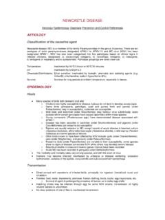
4 Other signs include fever, malaise, anorexia, rhinitis, sneezing, ptyalism and keratoconjunctivitis. Eighty per cent of recovered cats remain latently infected, and viral reactivation can cause recrudescent infections in 45% of these animals (Gaskell and Povey, 1977). Stromal involvement may occur as an immune response to the viral antigen. Upper respiratory signs are usually minimal or absent with recrudescent disease. FHV-1 disease syndromes include keratoconjunctivitis sicca (KCS), corneal sequestration, eosinophilic keratitis, ophthalmia neonatorum, symblepharon (Figure 2), uveitis and periocular dermatitis.
5 Symblepharon is an acquired conjunctival adhesion, which is rare in species other than cats. Primary disease caused by FHV-1 is common, but often self-limiting. Recrudescent disease is less common, but very frustrating. Feline calicivirus usually causes a less severe disease, with ocular signs usually limited to conjunctivitis, mucoid discharge and, sometimes, chemosis. Many cases have oral ulceration, and frequently there is a serous oculonasal discharge. Polyarthritis may be present. Chlamydiophila felis (formerly Chlamydia psittaci var. felis) can cause unilateral or bilateral conjunctivitis with marked chemosis and mucoid Figure 1: Kitten with FHV-1 infection, with typical conjunctivitis and nasal 2: Symblepharon the conjunctiva is adhered to the lateral cornea, with some 59 (4) : april, 2006 continuing educationirish Veterinary Journal 224to mucopurulent discharge.
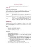
6 Corneal and upper respiratory tract involvements are not bronchiseptica causes a mild upper respiratory tract infection with oculonasal discharge and conjunctivitis. A severe form of the disease can occur, especially in combination with other upper respiratory tract pathogens, causing lymphadenopathy, sneezing, coughing and adventitious lung felis may be isolated from normal eyes, or it may cause a pseudomembranous conjunctivitis with conjunctival conjunctivitis is a not uncommon condition in older cats, which is often not noticed and has had little representation in the literature. Lipogranulomas are found distributed under the palpebral conjunctiva on upper and lower eyelids, and appear to be associated with the meibomian glands (Figure 3).
7 They present as single or, more commonly, multiple, raised, cream-white, smooth nodules within the palpebral conjunctiva. They may be present in one or more lids, and they are more commonly present in non-pigmented eyelids (Read and Lucas, 2001). The lesions may lead to chronic discomfort. nictitating membrane Usually, the normal Feline third eyelid is hidden, with the leading edge barely noticeable. Prominence of the third eyelid is noted in cats with a number of conditions. aetiology of unilateral and bilateral protrusions is presented in table 1. Interestingly, loss of the retrobulbar fat pad, debility and dehydration do not cause protrusion of the nictitating membrane to the same extent as they do in systemThe cat does not blink as frequently as the dog, and the corneal tear film is more stable.
8 However, keratoconjunctivitis sicca (KCS) can occur in cats and is commonly overlooked. Schirmer tear tests should be performed in cases of conjunctivitis or surface ocular disease. Normal values are 5mm to 11mm in one minute, but they need to be interpreted in conjunction with evidence of ocular surface disease. clinical presentation depends on the stage at which the disease is presented and varies, from conjunctival hyperaemia with superficial keratitis to severe ulcerative keratitis, with mucoid to mucopurulent discharge. Causes can be neurogenic ( , dysautonomia), drug induced ( , atropine administration) or idiopathic, or can be due to trauma, FHV-1, symblepharon or mucin abnormalities.
9 Apart from quantitative KCS, qualitative KCS can also cause corneal disease, thought to be due to a deficiency in tear mucin (Cullen et al., 1999)Figure 3: Lipogranulomatous conjunctivitis multiple white nodules in the palpebral conjunctiva of the lower 1: Protrusion of the third eyelid in cats unilateralglobe factorsthird eyelid factorsSympathetic innervation: Horner s syndromeSize: microphthalmosPosition: enophthalmos, , Horner s syndrome exophthalmos, , retrobulbar space- occupying lesionProlapse of the gland of the third eyelid (te)eversion of the cartilage of the teForeign body behind the teinflammation, , bulbar conjunctivitisSymblepharonneoplasiatraum aBilateralHaws syndrome with chronic diarrhoea (Figure 4)dysautonomia (Key-gaskell syndrome)drug-induced, , phenothiazine sedativestetanuscontinuing education Volume 59 (4) : april, 2006 irish Veterinary Journal 225corneaCorneal ulceration is commonly the result of trauma (Figure 5).
10 The position of the ulcer may give clues as to its aetiology . Central lesions may be due to exposure ( , facial nerve paralysis, exophthalmos, KCS, lagophthalmos). Perilimbal lesions may be caused by abnormalities of eyelids or cilia, while medial lesions may indicate a foreign body behind the third eyelid. All cases of conjunctival and ocular surface disease should be stained with fluorescein dye. There are frequently signs of unilateral ocular discomfort, such as blepharospasm, epiphora and, sometimes, photophobia. The corneal epithelial deficit may be very superficial (usually the most painful presentations) or it may involve the deeper stroma.