Transcription of Five Stages of Evolving -Cell Dysfunction During ...
1 five Stages of Evolving -Cell Dysfunction During progression to diabetes Gordon C. Weir and Susan Bonner-Weir This article proposes five Stages in the progression of diabetes then progress to the IFG or IGT range, where they diabetes , each of which is characterized by different may remain for years before developing frank diabetes . changes in -cell mass, phenotype, and function. stage 1 Although this progression is mostly discussed in the is compensation: insulin secretion increases to maintain context of type 2 diabetes , very similar changes occur as normoglycemia in the face of insulin resistance and/or type 1 diabetes unfolds and as pancreas or islet transplants decreasing -cell mass. This stage is characterized by fail. maintenance of differentiated function with intact We propose that there are five Stages in the progression acute glucose-stimulated insulin secretion (GSIS).
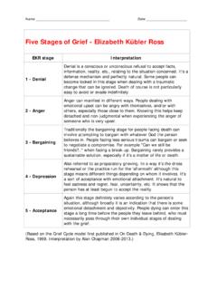
2 Of diabetes (Fig. 1), each of which is marked by important stage 2 occurs when glucose levels start to rise, reach- ing mmol/l; this is a stable state of -cell changes in -cell mass, phenotype, and function. Although adaptation with loss of -cell mass and disruption of conventionally thought of as a continuum, we make the function as evidenced by diminished GSIS and -cell case that each has its own important characteristics. stage dedifferentiation. stage 3 is a transient unstable period 1 is best described as compensation: insulin secretion of early decompensation in which glucose levels rise increases to maintain normal glucose levels in the face of relatively rapidly to the frank diabetes of stage 4, which insulin resistance resulting from obesity, physical inac- is characterized as stable decompensation with more tivity, and genetic predisposition.
3 stage 2 occurs when severe -cell dedifferentiation. Finally, stage 5 is char- glucose levels rise to levels of mmol/l (89 116. acterized by severe decompensation representing a pro- mg/dl) a stable state of -cell adaptation. stage 3 is an found reduction in -cell mass with progression to unstable period of early decompensation in which glucose ketosis. Movement across Stages 1 4 can be in either direction. For example, individuals with treated type 2. levels rise relatively rapidly to stage 4, which is character- diabetes can move from stage 4 to stage 1 or stage 2. For ized as stable decompensation. Finally, there is the severe type 1 diabetes , as remission develops, progression decompensation of stage 5 that represents profound -cell from stage 4 to stage 2 is typically found. Delineation of failure with progression to ketosis. Movement across these Stages provides insight into the pathophysiology Stages 1 4 can be in either direction.
4 For example, indi- of both progression and remission of diabetes . diabetes viduals with type 2 diabetes who undergo gastric reduc- 53 (Suppl. 3):S16 S210, 2004 tion surgery can move from stage 4 to stage 1. Even conventional treatments with diet, exercise, and oral agents often return people to stage 2. For type 1 diabetes , as remission develops, progression from stage 4 to stage 2. P. rogression to diabetes can be viewed as having is typically found. definable Stages characterized by changes in var- ious metabolic parameters and -cell function. At the very beginning, fasting plasma glucose levels stage 1: COMPENSATION. increase from perfectly normal values of mmol/l (80 The most common example of compensation is found with mg/dl) to higher values that might be as low as mmol/l the insulin resistance due to obesity, which is accompa- (89 mg/dl).
5 This change in glycemia would not be recog- nied by higher overall rates of insulin secretion (2) and nized as being clinically abnormal because it would fail to increased acute glucose-stimulated insulin secretion reach the official category of impaired fasting glucose (GSIS) following an intravenous glucose challenge (3). (IFG; glucose level mmol/l or 100 mg/dl) or impaired Much of the increase in insulin secretion undoubtedly glucose tolerance (IGT; 2-h postglucose level of results from an increase in -cell mass, as has been found mmol/l or 140 mg/dl) (1). Those destined to develop in autopsy studies in humans (4 7) and numerous rodent models (8,9). -Cell mass is normally tightly maintained From the Section on Islet Transplantation and Cell Biology, Joslin diabetes through a balance of -cell birth ( -cell replication and Center, Boston, Massachusetts.)
6 Islet neogenesis) and -cell death through apoptosis (10). Address correspondence and reprint requests to Gordon C. Weir, MD, Most of the increase in -cell mass with insulin resistance Section on Islet Transplantation and Cell Biology, Joslin diabetes Center, One Joslin Place, Boston, MA 02215. E-mail: is probably due to increased -cell number, but -cell Received for publication 10 March 2004 and accepted in revised form 31 hypertrophy may also contribute. It is not yet clear if the May 2004. This article is based on a presentation at a symposium. The symposium and higher plasma insulin levels can be entirely explained by the publication of this article were made possible by an unrestricted educa- the larger -cell mass or whether there is also increased tional grant from Servier. secretion per given unit of -cell mass. Although compen- GSIS, glucose-stimulated insulin secretion; IFG, impaired fasting glucose.
7 IGT, impaired glucose tolerance. sation is usually thought of in the situation of insulin 2004 by the American diabetes Association. resistance, similar changes presumably occur in the early S16 diabetes , VOL. 53, SUPPLEMENT 3, DECEMBER 2004. WEIR AND S. BONNER-WEIR. dinucleotide (NADH) to be oxidized by mitochondria, thereby contributing to adenosine triphosphate (ATP). formation. The oxidation of NADH should also enhance glycolytic flux. In - cells the malate/pyruvate shuttle may facilitate the generation of NADPH, which could somehow enhance secretion. The need for these shuttles probably explains why - cells have very high levels of mitochon- drial glycerol phosphate dehydrogenase (mGPDH) and pyruvate carboxylase (17,18). To maintain this degree of specialization, the genes that are highly expressed in - cells include those of the secre- tory products (insulin and islet amyloid polypeptide [IAPP]), key genes for glucose metabolism (GLUT2 and glucokinase), key enzymes for the mitochondrial shuttles (glycerol phosphate dehydrogenase [mGPDH]) and pyru- vate carboxylase, and critical transcription factors (PDX-1.)
8 And ). Yet, enzymes that participate in unwanted pathways such as gluconeogenesis and lactate production FIG. 1. Schema of five Stages of progression of diabetes . are expected to be suppressed. Some of these include phosphoenolpyruvate carboxykinase (PEPCK), glucose-6- Stages of autoimmune destruction. As -cell mass falls, phosphatase and fructose-1,6-bisphosphatase, and lactate there must be a signal to increase mass and secretion, dehydrogenase. Hexokinase would also be unneeded, so which presumably prolong the prediabetic period, which its expression could also be suppressed. Clearly these can last for years (11). changes in gene expression are just the tip of the There is much interest in the signal leading to increased iceberg, and a full picture of -cell phenotype awaits -cell mass in this situation. The facile, but probably gene array studies, which should emerge in the near correct, explanation is that there is a feedback loop with future.
9 Insulin resistance producing increased glucose levels that Maintenance of -cell differentiation During stage 1. stimulate -cell secretion and growth. However, concerns compensation. Some experimental data support the con- are raised about how this can be compatible with seem- cept that much of the -cell phenotype is kept intact ingly normal glucose levels. One explanation is that the During successful compensation for insulin resistance. feedback loop is tightly regulated, like a thermostat that When glucose was infused into rats for 4 days, compensa- can maintain temperatures within a very narrow range. A tion occurred and glucose levels remained normal During molecular mechanism that could facilitate this control is the latter Stages of the infusion because of increases in activation of glucokinase in - cells , the enzyme that con- insulin secretion that were accompanied by increased trols the rate of glycolysis, thereby determining insulin -cell mass (19).
10 While responding to the increased de- secretory rates (12). Thus, very small elevations in glucose mand, the -cell phenotype, as determined by analysis of a levels could lead to a change in the set point in GSIS that selected group of genes, remained remarkably similar to allows maintenance of normal plasma glucose levels the control profile (19). There must have been induction of (13). some genes required for -cell replication, but the genes The importance of -cell differentiation. The unique responsible for maintaining the machinery allowing GSIS. differentiation of - cells is responsible for the extraordi- did not seem to be perturbed. This preservation of pheno- nary efficiency of these cells in storing and secreting type, coupled with an increase in -cell mass, is consistent insulin to provide precise regulation of metabolism. With with the exuberant GSIS seen in obesity.