Transcription of FLATFOOT Definition - Bonefix
1 FLATFOOT Definition Flat foot is defined as a condition in which there is a loss of medial longitudinal arch on weight bearing. It can be quantified by means of footprints 23% in normal population (Harris and Beath) have FLATFOOT Flat foot can be associated: Hind foot valgus Achilles contracture Genu Valgum Greater prevalence of flatfeet in shod than in unshod kids in india Types I. Flexible or Hypermobile FLATFOOT with normal Achilles 65% of FLATFOOT and 15% all population No clinical concern Benign and normal variation Look for: Hyperlaxity [Marfans, Downs] Accessory navicular II. Flexible flat foot with tight Achilles 25% of FLATFOOT and 7% of normal population Associated contracture of achilles and may be symptomatic. Normal excursion of subtalar joint III. Rigid flat foot 10% of FLATFOOT [3%] Causes: Subtalar arthritis Tarsal coalition Subtalar infection Old fracture Vertical talus IV Neuromuscular Cerebral Palsy Spina bifida V Adult onset FLATFOOT Tibialis posterior dysfunction Assessment 1.
2 Family history 2 Look for abnormal callosity 3. Look for ligament laxity 4. Check the shoe for wear pattern 5. Foot print 6. Hindfoot movement 7. Look for tendo achilles contracture The talonavicular joint is locked in inversion Dorsiflexion of the foot with knee in extension and flexion. 8. Jack test: Patient weight bearing. On dorsiflexing the great toe, the arch of the foot exaggerates in a flexible FLATFOOT . 9. On standing tip toe: In flexible FLATFOOT , arch appears indicating that the hindfoot is flexible. If there is no change in the heel on standing tip toe, the FLATFOOT is rigid 10. Test for Tibialis posterior dysfunctional syndrome 11. Neurological assessment X RAY Weight bearing radiographs 1. Lateral view: Look for a sag at talonavicular or Naviculo cunieform joint 2. Talo Calcaneal angle AP: Talo Calcaneal angle: 15 to 35 ; >35 valgus heel Lat: Talo Calcaneal angle: 25 50 3.
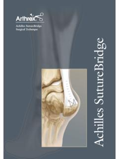
3 Talo Metatarsal angle:[Meary] 0 is normal In Cavus foot 0 to 15 is a mild deformity 15 to 30 is moderate > 30 is severe. In Planovalgus feet, this angle is negative Illustrations of usefully measured angles. A: AP views demonstrating TC = talocalcaneal and T 1stMT = talo first metatarsal angles. B: Lateral views demonstrating A = tibiotalar; B = talo first metatarsal; C = talohorizontal; D = talocalcaneal; and E = tibiocalcaneal angles Treatment 1. In most an arch will develop by 5 years. In children, reassure the parents and explain the benign nature. 2. Exercise, shoe inserts and modification are ineffective 3. In symptomatic patient [pain] or severe shoe wear consider orthotic support. Medial arch support and heel cup 3. When heel cord tightness is present Suggested stretching exercises Rarely heel cord lengthening or lengthening of lateral column.
4 4. Rare bony surgeries for flat foot Calcaneal osteotomy Miller fusion: Navicular I Cuneiform I metatarsal Lateral column lengthening THE LATERAL COLUMN LENGTHENING A bone graft from the tibia is inserted into the anterior process of the calcaneum. This procedure preserves the calcaneo cuboid joint and pushes the navicular bone medially. The osteotomy is between the anterior and middle facet of the calcaneum MEDIAL DISPLACEMENT CALCANEAL OSTEOTOMY Make a lateral oblique incision. Retract the peronie tendons superiorly Osteotomise the calcaneus perpendicular to the body between the posterior facet and the tuberosity. Open the osteotomy site with a lamina spreader Now displace the medial calcaneal tuberosity medial cannulated screw CONCLUSIONS 1. No long term prospective study regarding natural history.
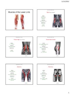
5 2. Prognosis is good 3. Longitudinal arch in the child's foot develops spontaneously during the first 5 year 4. When Achilles contracture: need stretching and sometimes tendo achilles lengthening 5. Arch supports has not shown to improve the height of the arch 6. Severe: Soft tissue procedures or combine osteotomies and arthrodesis to restore longitudinal arch 8. CT: Rule out any coalition SKEW FOOT Rare; Genetic Hindfoot deformity of FLATFOOT and forefoot adductus Etio: Idiopathic/Iatrogenic X ray: Skewfoot: An AP radiograph reveals the significant varus deformity of the forefoot creating a negatively valued talo first metatarsal angle. Delayed ossification of the navicular prevents radiographic documentation of the lateral talonavicular subluxation. The hindfoot valgus is portrayed by the increased talocalcaneal angle Treatment Symptomatic Evans Calcaneal lengthening osteotomy And Medial cuneiform opening wedge osteotomy and lengthening of Achilles CONGENITAL VERTICAL TALUS Talus is in plantar flexion with dorsal dislocation of the navicular on the talar head and neck.
6 Vertical position of the talus is seen in X rays in both dorsiflexion as well as in plantarflexion. In severe form may be associated with Calcaneo cuboid joint involvement. Etiology 1. Defects of CNS: Myelogmeningocele Diastematomyelia Sacral agenesis Arthrogryposis Cerebral palsy, Polio 2. Iatrogenic: Overcorrection of a clubfoot 3. Idiopathic 4. Genetic: Patau syndrome trisomies of 13 15, Edwards syndrome Whistling face and prune belly syndrome Pathology Bones Talus and calcaneus plantarflexed Navicular dorsiflexed Ligaments Stretched ligament: Spring ligament, Talo calcaneo navicular ligament Anterior fibers of Deltoid Contracted ligament: Lateral portions of dorsal talonavicular, Interosseous talo calcaneal ligament Muscles Contracted: Achilles, Tibialis Anterior, EHL, Peroneus longus and brevis Lengthened: Tibialis Posterior, FHL, FDL Retinaculum Dorsal retinaculum thickens Peroneal retinaculum is lax and allows subluxation of the tendon Assessment 1.
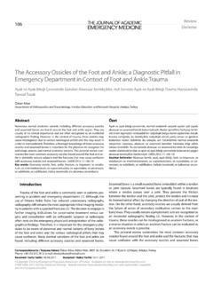
7 Persian slipper foot appearance 2; Peg like gait (walks on the talar head) 3. Deformities in rocker bottom foot a) Hindfoot : Equinovalgus b) Midfoot valgus c) Dorsal dislocation of Talo navicular joint d) Forefoot abduction 4. Neurological assessment 5. Hip and spine assessment X ray Lateral foot in Dorsiflexion and plantar flexion Vertical Vs oblique Talus Calcaneal pitch angle is low Vertical Oblique Vertical Talus: An AP radiograph demonstrates an increased talocalcaneal angle due to the equinovalgus angulation of the os calcis. The lateral view depicts the significant hindfoot equinus and vertical position of the talus. Thus, the talohorizontal and tibiotalar angles approach 90 and 180 , respectively. The maximum dorsiflexion view reveals the rigidly fixed hindfoot equinus. Oblique talus: Comparison of the maximum dorsiflexion and plantar flexion views demonstrate the reduction of the midfoot on the hindfoot in plantar flexion which establishes the diagnosis of oblique talus.
8 MRI is useful as it can outline the cartilaginous analogue of the bones. MRI of spine when spinal pathology is suspected Treatment 1. Patient with neurological problem have less favourable functional outcomes and may require talectomy if recurrence of deformity following soft tissue release 2. In majority the procedure is performed between 6 12 months Principle of surgical treatment Cincinnati incision The heel cord is lengthened and Posterior tibial Neurovascular bundle is identified FHL, FDL and Tibialis Posterior is divided Posterior capsulotomies Lateral subtalar capsulotomy and divide contracted Calcaneo fibular ligament Capsulotomy of Calcaneo Cuboid joint Z lengthening Peronie, EHL and Tibialis anterior Shorten: FHL, FDL, Tibialis posterior 3 wires: Talo Calcaneal; Calcaneo Cuboid; Talo Navicular Differential diagnosis Metatarsus Adductus: An AP radiograph demonstrates varus deformity of the forefoot relative to the hindfoot as represented by the negative value of the talo first metatarsal angle.
9 The talocalcaneal angle is normal. Skewfoot: An AP radiograph reveals the significant varus deformity of the forefoot creating a negatively valued talo first metatarsal angle. Delayed ossification of the navicular prevents radiographic documentation of the lateral talonavicular subluxation. The hindfoot valgus is portrayed by the increased talocalcaneal angle (the upper limits of normal as shown). Clubfoot: A: On the AP radiograph, the "parallelism" of the talus and calcaneus and negatively valued talus first metatarsal angle demonstrate the varus deformities of both the hindfoot and forefoot, respectively. The lateral view reveals the decreased talocalcaneal angle characteristic of the hindfoot equinovarus deformity. Moderate stacking of the metatarsals depicts the forefoot supination. Tarsal coalition: The 45 internal oblique view may prove useful for diagnosis of calcaneonavicular coalitions.
10 The lateral view may demonstrate the "anteater nose sign" typically characteristic of calcaneonavicular coalitions. Note the tubular elongation of the anterior aspect of the calcaneus towards the mid navicular region. Conclusion Radiographs are a critical tool in evaluation of pediatric foot abnormalities. The resulting angular measurements can define the boundaries of normal and assist in the diagnosis as well as quantify the degree of deformity. However, accurate and reproducible radiographic studies are essential for effective use of these lines and angles. Small flaws in radiographic techniques may significantly alter these measurements. Ultimately, diagnosis of congenital foot deformities is accomplished clinically. Radiographic studies are an effective adjunct used to exclude unexpected deformities or to document the evolution of deformities.