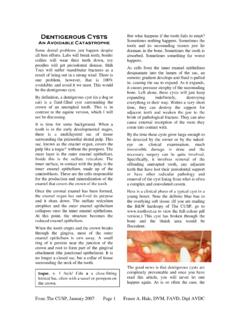
Transcription of Fraser Hale, DVM, FAVD, DipAVDC - Hale …
1 pulp Exposure. F. A Hale, DVM, favd , DiplAVDC, 2011 Fraser Hale, DVM, favd , DipAVDC This informational page is an attempt to describe a complex subject in limited space. It should be considered to be very introductory. More in-depth discussions of this and many other subjects can be found on the Old CUSP Articles page at You are encouraged to visit and make use of the resources there and elsewhere on my website. pulp Exposure in Dogs and CatsDog and cat (and your) teeth are made up of four tissues. The crown is covered by a thin layer of very hard, non-porous enamel. The enamel all forms before the tooth erupts to become visible in the mouth.
2 The root is covered by a thin layer of cementum. Under the enamel of the crown and cementum of the root is the tissue that makes up the bulk of the mature tooth called dentin. It is harder than bone but not as hard as enamel. When the tooth first erupts, the dentin wall is very thin and dentin is laid down on the inside of the tooth so over time, the dentin wall gets thicker. In the hollow space inside the tooth is the dental pulp . pulp contains the cells that produce dentin inside the tooth, blood vessels, nerves and various other tissues. The pulp is very sensitive with a high concentration of pain receptors.
3 Normally, the only way in or out of the pulp chamber is through a collection of tiny channels at the tip of the roots (known as the apical delta). When the crown of a tooth is damaged (worn down or fractured) in a way that exposes the pulp tissue inside the tooth, it puts into motion an inevitable series of events. Once the pulp tissue is exposed to the oral environment, it will become contaminated with oral bacteria. It will then become infected and inflamed. In time the infection will kill the pulp tissue and then the infection can spread out through the root tips into the space between the root and bone and then into the bone itself.
4 All of this is very painful (toothache). Once the pulp is dead the tooth can be viewed as a hollow tube embedded in the bone into which the animal is spitting every day. It is an open pathway for infection that the body has no way of closing. This series of events (contamination of the pulp , inflammation of the pulp , septic necrosis of the pulp , infection in the area around the root tips) will happen 100% of the time when a tooth suffers a pulp exposure. How long a tooth takes to go through all those stages varies, but it will happen to all of unless appropriate treatment is instituted.
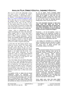
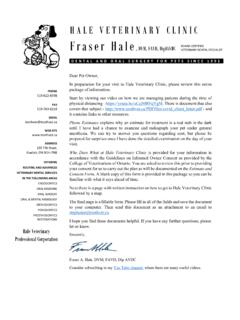
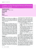
5 What is appropriate treatment? In general, there are two options root canal treatment of some form (total pulpectomy or vital pulp therapy) or extraction. Sometimes the damage is too severe to consider root canal treatment in which case extraction is the only option. Teeth with pulp exposure are not a wait-and-see proposition. They cannot be saved or managed with antibiotics and they cannot seal themselves . They absolutely need root canal treatment or extraction. Left: Pre-op radiograph of a fractured lower right 1st molar with septic pulp necrosis and infection in bone around the root tips.
6 Right: radiograph of this same tooth one year after root canal treatment showing healing of the bone. BOARD-CERTIFIED VETERINARY DENTAL SPECIALIST DENTAL AND ORAL SURGERY FOR PETS Diagram and radiograph of a dogs lower right 1st molar tooth.