Transcription of G E N E R A L Tutorial 317 - AAGBI
1 GENERAL Tutorial 317. UNDERSTANDING and ESTABLISHING. INTRAOSSEOUS ACCESS. Dr. Scott Bradburn ST3 Anaesthetic Registrar, Glangwilli General Hospital, Wales Dr. Stuart Gill Anaesthetic Consultant, Glangwilli General Hospital, Wales Edited by Dr. Matthew Doane 26th JUNE 2015. Correspondence to QUESTIONS. Before continuing, try to answer the following questions. The answers can be found at the end of the article, together with an explanation. Please answer True or False: 1. Regarding humeral IO access: a. Infusion rates comparable to those through subclavian central lines can be achieved b. Can be used to safely administer resuscitation drugs c. Landmarks for IO insertion points are always easy to find in obese patients d. It is the most painful site for insertion 2.
2 The following are suitable sites for IO access in young children: a. Distal femur b. Sternum c. Proximal tibia d. Humeral head 3. The following are contraindications for IO access: a. Fracture proximal to the proposed IO insertion point b. Burns distal to the proposed IO insertion point c. Local sepsis over proposed IO insertion site d. Systemic sepsis 4. The following are complications of IO access: a. Extravasation b. Microfracture c. Osteomyelitis d. Osteoporosis Key Points INTRODUCTION. The intraosseous (IO) route is an underutilised, Intraosseous (IO) access is an alternative method to reliable and extremely valuable alternative to providing venous administration of drugs and fluids. venous access Commonly used by the military and pre-hospital medics, intraosseous access has expanded its use IO access can be used in trauma, emergencies, to a variety of settings: in the emergency department, and in cases requiring volume resuscitation at cardiac arrests, in the paediatric population, and is gaining popularity in adult settings where intravenous There are several devices and sites that may be access is challenging or time critical.
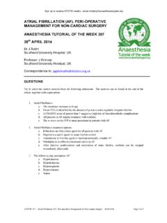
3 Used for IO access The IO route is now endorsed by The Resuscitation Inserting an IO needle is easy to learn, but Council UK and is in the current Adult Life Support success depends on familiarity with the (ALS) and Advanced Paediatric Life Support (APLS). equipment and correct technique, as described guidelines (1,2).. in this Tutorial Subscribe to ATOTW tutorials by visiting ATOTW 317 Understanding and Establishing Intraosseous Access (26th June 2015) Page 1 of 8. VENOUS BLOOD DRAINAGE FROM BONES. Theoretically, intraosseous access can be obtained in any large bone and current devices support several specific points of access, including the sternum. As bones are non-compressible, the intraosseous space will stay patent, even in shocked patients.
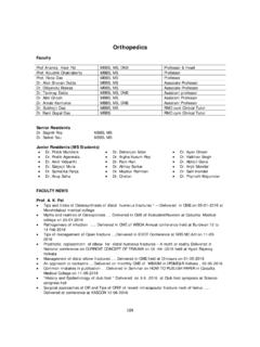
4 This provides a readily available route for infusion of drugs or fluids in an emergency, while also providing access to bone marrow (3,4). aspirate, which can be used for some simple blood tests . The venous plexus of long bones has been shown to drain in to the central circulation, at a rate comparable to central (4-7). venous access . (8). Fluid resuscitation can also be accomplished via the IO route , with respectable flow rates of 1-3L/hour via tibial access or 5L/hour via humeral access. Due to the intrinsic pressure of the intraosseous space, infusions commonly do not flow effectively with gravity alone and need to be administered under pressure using pressure bags, syringe driver or manual flushing. FIGURE 1: Vascular anatomy of long bones.
5 Intraosseous access is achieved in the trabeculated space of the bone. (Courtesy of Vidacare/Teleflex). INDICATIONS. There are numerous conditions where IV access may be difficult and this can be overcome by using the intraosseous route. All forms of physiological shock, hypothermia, multiple previous intravenous lines, or intravenous drug use are (9-12). common situations where IO access has proven invaluable . CONTRAINDICATIONS. Absolute Bone trauma at or proximal to the insertion site, or previous IO insertion in the same limb: disruption of the bone at or proximal to the site allows for extravasation of infusions and potentially the development of (9-11). compartment syndrome . Infection overlying the point of insertion: there are concerns of seeding the infection into the bone and (11-13).
6 Causing osteomyelitis Relative Prosthesis in the target limb (knee replacement, tibial nail, humeral plate), or previous sternotomy: the disruption of the bone matrix can unpredictably interfere with insertion or flow rates and insertion into in-dwelling metal work could potentially cause damage to the prosthesis or IO needle. Difficulty in identifying anatomical landmarks: in these patients, IO devices should be deployed with (12). extreme caution, as damage could be caused to other underlying structures . Subscribe to ATOTW tutorials by visiting ATOTW 317 Understanding and Establishing Intraosseous Access (26th June 2015) Page 2 of 8. COMMON ACCESS SITES. While there are several insertion points described, it is important to acknowledge that some devices are limited to specific anatomical sites.
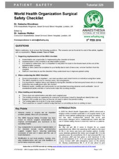
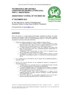
7 The anatomical landmarks for the most common sites are described below: Humeral head With the elbow bent, and the patient's hand on their abdomen, palpate the surgical neck of the humerus to locate the greater tuberosity. The insertion site is approximately 1 cm above the surgical neck and 2-3cm lateral to the biceps tendon (Figure 2). Insert at a 45-degree angle, aiming towards the opposite scapula. The manufacturer does not (11). recommend this technique in paediatric patients, but clinical judgement will need to be used . FIGURE 2: Landmarks for humeral insertion (Image courtesy of Vidacare). Proximal tibia 2cm medial and 1-2 cm below the patella, palpate the tibial tuberosity and ensure that you can feel bone below subcutaneous tissue (Figure 3).
8 The needle should be angled slightly distally, away from the knee(11). FIGURE 3: Landmarks for tibial insertion (Image courtesy of Vidacare). The following routes are less commonly used but may be considered if the proximal tibia or humerus are not available: Distal tibia Palpate the most prominent aspect of the medial malleolus, as well as the anterior and posterior borders of the tibia. (11). Ensure the flat part of the bone is located and insert 3cm proximally to this point, at 90-degrees to the skin . Distal femur With the leg fully extended at the knee, palpate the external condyles of the distal femur. The ideal insertion point is 2- 3cm superior and 1-2cm medial to the anterior midline (proximal placement is important to avoid growth plates in younger patients).
9 The leg should be immobilized until the cannulae is removed as movement at the ipsilateral knee may (16). cause the quadriceps tendon to displace the cannula . Sternum This route of insertion has been useful in military casualties, as the sternum is often protected by body armour and thus intact after major trauma. Locate the manubrium, approximately 2cm below the sternal notch, and insert at 90 degrees to (7,14). the skin. Fluid flows into the internal mammary veins, into the azygous veins, and then into the central circulation . Subscribe to ATOTW tutorials by visiting ATOTW 317 Understanding and Establishing Intraosseous Access (26th June 2015) Page 3 of 8. GENERAL ADVICE FOR ESTABLISHING IO ACCESS: In all IO access attempts, the following key points should be followed: 1.
10 Sterilisation of the skin at the needle insertion site 2. Manual stabilisation of the bone during insertion 3. Aspiration after needle insertion confirms successful placement 4. In the awake patient, injection of local anaesthetic (preferably lidocaine) into the IO needle prior to use can reduce pain for subsequent infusions 5. Ensure the needle is flushed with at least 10ml of fluid after drug administration 6. Clear documentation of the procedure in the patient notes 7. Frequent assessment of the IO site for signs of extravasation TYPES OF DEVICES. Manual trocar These require significant insertion force and are commonly used in paediatric patients for lower limb access sites. The main advantage lies in the simplicity of the device and lower cost.