Transcription of Genetics of Childhood Disorders: L. Learning and …
1 Described initially at the beginning of the 20th century byIvan Pavlov, classical conditioning involves the pairing of neu-tral stimuli with aversive or appetitive cues. This results in learn-ing. Formally neutral stimuli now predict salient events. Oneform of Pavlovian conditioning is fear conditioning, the pri-mary associative Learning mechanism involved in aversive emo-tional Learning . It is through this process that we learn to befearful of people, animals, objects, and places. From a psy-chological perspective, it is one of best understood types oflearning in mammals because the stimulus and response prop-erties can be very carefully controlled. For these reasons, Pavlovianfear conditioning has served as a powerful animal model of fearand anxiety disorders including phobia, panic disorder, post-traumatic stress disorder, and possibly many of the neurotic 612J.
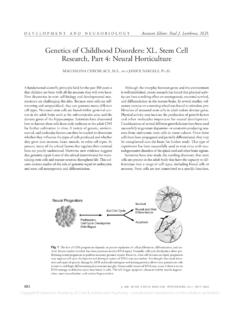
2 AM. ACAD. CHILD ADOLESC. PSYCHIATRY, 42:5, MAY 2003 DEVELOPMENT AND NEUROBIOLOGYA ssistant Editor: Paul J. Lombroso, of Childhood Disorders: L. Learning andMemory, Part 3: Fear ConditioningKERRY RESSLER, , , ANDMICHAEL DAVIS, 1(A) Pavlovian fear conditioning and measurement of fear-potentiated startle. Top: during training, the CS (light) is paired with the US (foot-shock). Duringtesting, the acoustic startle reflex is elicited in the absence (middle) or presence (lower) of the CS. Fear-potentiated startle is defined by higher acoustic startle ampli-tude in the presence of the fear-conditioned CS. (B) Schematic diagram illustrating the convergent inputs of US (pain) pathways and CS (multimodal sensory)pathways into the basolateral nucleus of the amygdala (BLA).
3 Neurons in the basolateral nucleus project to the central nucleus (CeA). Neurons in the centralnucleus project divergently to numerous brain regions which together mediate the fear response. CS = conditioned stimulus; US = unconditioned stimulus; VTA =ventral tegmental area; LC = locus ceruleus. (C) Schematic diagram illustrating some of the molecules implicated in the formation of fear memory. In brief, N-methyl-D-aspartate (NMDA) and other receptor activation leads to downstream intracellular events, eventually culminating in the transcription of numerous new genesinvolved in long-term memory consolidation. NMDAR = the N-methyl-D-aspartate glutamate receptor; AMPAR = the glutamate receptor which mediates celldepolarization; VGCC = voltage-gated calcium channel; TrkB = the tyrosine kinase receptor for brain-derived neurotrophic factor (BDNF); CaMKII = calciumcalmodulin-dependent protein kinase II; MAPK = mitogen activated protein kinase; AKAP = A-kinase anchoring protein; PKA = cyclic AMP-dependent proteinkinase; PKC = protein kinase C; PI-3K = phosphatidylinositol-dependent kinase; CREB = cyclic AMP response element binding of everyday life.
4 If we can understand the under-lying molecular basis for how a previously innocuous stimu-lus leads to intense fear in an animal model, the hope is thatsuch an understanding will eventually lead to better treatmentsin humans disabled by these crippling the obvious usefulness of this model system forunderstanding fear and anxiety disorders, fear conditioninghas received considerably more attention in recent years fortwo reasons. Investigators are able to carefully control the stim-ulus-response properties of fear conditioning in rodents. Thishas led to very precise behavioral paradigms for studying learn-ing and memory. In addition, the ability to combine behav-ioral, anatomical, and electrophysiological techniques, both invitro and in vivo, has contributed to an advanced understandingof the neural circuitry underlying this form of Learning .
5 Thiscombination of approaches has made fear conditioning andits neural substrates among the best models for the under-standing of Learning and memory across the cellular, neuralsystems, and organismal levels of organization. In this briefreview, we will first describe the experimental paradigm involvedin generating and measuring fear-conditioned Learning . Wewill then discuss the neural circuits and their cellular substratesthat are thought to mediate the Learning and memory general, fear conditioning involves the pairing of a pre-viously neutral cue, which then becomes the conditioned stim-ulus (CS), with an aversive cue, the unconditioned stimulus(US). In rodents, the US is typically given in the form of a mildfoot-shock (Fig.)
6 1A). Although any form of sensory cue maybe used, the CS is typically a discrete tone, light, or odor. Infact, rodents can easily discriminate among numerous discretecues in this form of conditioning. After several pairings of theCS and US, the animal will display a conditioned response tothe CS alone which is indicative of a central state of fear. Anytime after this training, from hours to months, a fear responsecan be elicited by exposure to the CS most commonly used measures of conditioned fear arethe freezing response and the fear-potentiated acoustic startlereflex. With both of these measures, the animal is placed in aneutral environment and the CS is presented in the absence ofthe US. Freezing is measured as immobility during the pre-sentation of the CS.
7 The percentage of time spent freezing isfound to be significantly greater in the presence than in theabsence of a fear-conditioned CS. Alternatively, the acousticstartle reflex is elicited by presenting the animal with a shortnoise burst which serves to elicit a reproducible startle response(Fig. 1A). Measurement of the startle reflex can be entirelyautomated such that a computer may be used to record themagnitude of the startle response as well as to control the pre-sentations of the CS and US. In the presence of the CS ( ,light, odor), the magnitude of the startle elicited by the noiseburst is reproducibly greater than it is in the absence of the CS,and this difference is referred to as fear-potentiated amygdala is the primary brain region involved in fear-conditioned Learning .
8 However, the amygdala in fact refers toa number of functionally separate nuclei that are anatomicallygrouped together. The learned state of fear involves activation ofthe basolateral nucleus of the amygdala which in turn activatesthe central nucleus of the amygdala (Fig. 1B). As its name implies,the central nucleus is a hub that serves to initiate the full fearresponse whether it is activated physiologically via the basolateralnucleus or experimentally through electrical fear response is generated through the hardwired neuralconnections that exist between the central nucleus and a num-ber of other neural pathways. For example, activation of vari-ous midbrain nuclei by the central amygdala results in freezing,potentiation of reflexes such as the acoustic startle reflex, andincreased respiration.
9 Projections to the lateral hypothalamusactivate the sympathetic nervous system leading to cardiovas-cular effects, pupil dilation, and increased sweating. Activationof the paraventricular nucleus of the hypothalamus activatesthe glucocorticoid response. Lesions of these individual brainregions that are downstream of the central nucleus serve to blockspecific aspects of the fear response, whereas ablation of the cen-tral nucleus itself blocks the entire fear addition to the well-understood circuitry underlying theoutputs of fear response, the sensory inputs representing theCS and US pathways have also been studied. Auditory, visual,and somatosensory projections arrive at the basolateral amyg-dala (BLA) through both thalamic and cortical pathways, on the other hand, take a more direct routefrom the olfactory bulb through the piriform cortex and directlyinto the amygdala.
10 In all cases, the pathways representing theaversive US and the previously neutral CS converge in the is within the BLA that the most critical cellular processesunderlying associative Learning are thought to is a large amount of data supporting a role for the BLAin initiating associative fear Learning . Chemical and electricallesions of the BLA have been shown to block new fear learn-ing. Furthermore, the principal electrophysiological measure oflearning, long-term potentiation (LTP), appears to occur betweenthe neurons projecting to and the target neurons within theBLA in brain slices. Electrical stimulation of neurons withinthe BLA before and after fear conditioning in freely moving ratshas shown that there is potentiation of the pathway mediatingmany of the sensory connections to neurons within the together, these data suggest that fear conditioning mayresult in an alteration in the functional connectivity betweensensory pathways and the BLA, such that future presentationof the CS alone is now sufficient to activate the central model for the circuitry changes underlying fear con-ditioning proposes that when the associative CS-US pairingoccurs, strong firing of neuronal projections that mediate theaversive stimuli are paired with weaker firing representing theneutral stimuli.