Transcription of Hematology Essentials: A Foundation for Accurate …
1 Christine Hinz, MS, MLS(ASCP)CMHematology Essentials: A Foundation for Accurate smear Reviews Current Challenges System Wide Approach StandardizationDifferential Training ProgramTraining AreaHow does the program work? Training Material Trainer and Trainee Checklists Reference guides Actual patient slides Case Study Power Point Competency Checklist I have to tell you I was dreading the diff training BUT it was AWESOME! Where I worked before I didn't have any formal diff training, you knew the basics and that was it. I love all the hand outs you provided. Things made more sense after the class. Trainee Feedback I was worried about the time commitment, but every employee came back saying how valuable the training I was impressed to hear how excited my employees were about Hematology after the training Employees feel they really have the tools now to provide great patient care Manager Feedback Follow up post training with Cellavision images for competency Ongoing competency assessment Adapted for smaller sites and/or affiliatesPost Training smooth.
2 Homogenous film 1/2 to 3/4 the slide length straight feather edge at least 1/4 inch examination area pink RBCs and appropriate WBC blues under gross examination(Rainbow feather edge)Proper Slide PreparationBad slide prepGood slide prepThe Good and Bad Examine on 10X: Check for good cell distribution, free of precipitate Examine extreme feather edge: Platelet clumps Look for abnormal cells: More dense and larger cells will be pushed to the feather edgeStarting your slide examination Area between extreme feather edge and Zone of Morphology is the cobblestone area. DON T do the morph or diff in this area. Zone of Morphology -area where cells evenly distributed, RBC s close but not touching.
3 Diff and morphology should be performed hereStarting your slide examinationZone of morphology Make sure slide has been made correctly If the slide has been pushed too hard when making the slide, WBC s will be concentrated at extreme feather edge and estimate will not match instrument result. WBC Estimate Estimate the white count under 10x or 40X/50x. Under low power 10X: 5 WBC's = 1,000/cumm Under 40X/50X: 1 WBC = 2,500/cumm The white count estimate may not be reported, but every manual differential white count is checked in this mannerWBC EstimateIn Zone of Morphology : Switch to 40x/50X or 100X to count 100 WBC cells.
4 Note: Perception at 100x can be distorted Manual differential vs analyzer differential Must drop to 100X for RBC morphology and Platelet estimate. Platelet Estimate = (Total # of PLTs Counted in 10 Fields Using 100X ) X 15,000 Performing a manual differential Morphology not reported: Anisocytosis, Macrocytosis, Microcytosis, Poikilocytosis, Stomatocytes Morphology reported as present: Toxic Granulation, DohleBodies, Auer Rods, HypersegmentedNeutrophils, HyposegmentedNeutrophils, Vacuolated Neutrophils, Reactive Lymphocytes, Smudge Cells, Large Platelets, AgranularPlatelets, Dwarf Megakaryocytes, Atypical Platelets, BasophillicStippling, PappenheimerBodies, Howell Jolly Bodies, Sickle Cells, RouleauxKnow appropriate morphology reporting Slight, Moderate, Marked: Hypochromasia, Polychromasia Few, Moderate, Many: Target, Acanthocytes, Echinocytes, Schistocytes, Spherocytes Platelet estimate choices.
5 Decreased, Adequate, Increased, ClumpedKnow appropriate morphology reportingReview Blood Maturation Chart N/C Ratio Chromatin pattern-clumped or fine Nucleoli Cytoplasm-Color of granules, inclusions Size of cell Myeloid Series-5 characteristics to look for PMN-coarse chromatin Lymph-N/C ratio 5:1 to 2:1 chromatin pattern clumped. Sky blue cytoplasm Large Lymphnucleus off center/clear cytoplasm (size determined by type of lymph, B,T,Killer) Basophil-large purple granules-see increase in reactive conditions such as MPD(myloproliferativedisease.) Monocyte Eosinophil-contain bright orange-red granules evenly distributed in the cytoplasm-rarely overlie the nucleus.
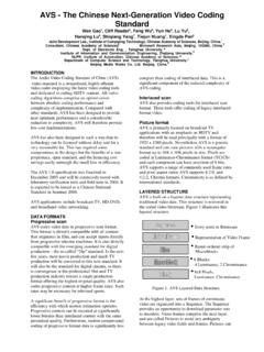
6 Band-narrowing of nucleus by 50%Normal SlideCopyright 2002 American Society of Hematology . Copyright restrictions may , P. ASH Image Bank 2002;2002:100360 NeutrophilLymphocyte7-16 m, nucleus is the size of a normal RBC, condensed chromatin, granules may be presentMonocyte12-20 m, folded nucleus, lacy chromatin, blue-gray cytoplasm, fine and Basophil12-15 m, 2-3 lobed nucleus, prominent reddish-orange granules10-15 m, segmented nucleus, prominent blue granulesSlides courtesy of Neutrophil9-15 m, horseshoe shaped nucleus, chromatin present in any filaments Leukemia is the uncontrollable growth of cells. Demonstrates a variety of immature cells, including blasts Basophiliaand a left shift can be some of the first signs of CML Cells to be identified on slide.
7 Myelocyte Metamylocyte-Nucleus kidney bean shaped Promyelocyte-(granules can overlap nucleus) Basophilic cytoplasm-Chromatin pattern is fine 1-2 nucleoli NRBC Myeloblast-Most immature cell in the myeloid series, N/C ratio high-fine chromatin pattern, basophilic cytoplasm Chronic Myeloid LeukemiaCML Mononuclear cells seen on slide Not seeing RBC s overlapping on slide Not seeing many platelets Pancytopenia-All three cell lines are affected Don t see many neutrophils (neutropenia) Large lymphs(clear cytoplasm/offset Nucleus) Blasts: Note-If you see Auer Rods this indicates cell is in the myeloid lineageAcute Myeloid Leukemia RBC morphology sometimes seen on slide.
8 Basophilic stippling Polychromatic Elliptocytes(Ovalocytes) Teardrops NRBCsAcute Myeloid LeukemiaAMLAMLB last vs Lymph 4yr old, cough, fatigue High WBC count, low , low Plt-20,000 Mononuclear cells with high N/C ratio, fine very fine, smooth chromatin pattern Slide full of BlastsALLALL Affects B-cell lymphocytes Typical Lymphocytosis > absolute Characteristic nucleus that looks like cracked earth or a soccer ball Cells are fragile, resulting in smudge cells present on smear Albumin slides made to reduce smudge cells, diff should be performed on albumin slide, RBC/WBC morphology should be performed on the original slideCLLCLLCLL vs ALL Variability of cellular size and shape as well as nuclear size, shape and chromatin pattern Seen in many viral illnesses-infectious mononucleosis Nucleus attached to cell wall Cytoplasm surrounding RBC s Reactive lymph vs MonocyteReactive LymphsReactive LymphsReactive Lymph Used to boost WBC following chemo Toxic granulation DohleBodies-sometimes Immature cellsGCSF.
9 Neulasta, Neupogen Toxic Granulation-Large, purple or dark blue azurophilicgranules, resembling the primary granules of promyelocytes, in the cytoplasm of neutrophils, bands and metamylocytes. Seen in severe infection, chemical poisoning, and other toxic states DohleBodies-Appear as single or multiple light blue or gray staining area in the cytoplasm of neutrophil. RNA and represent failure of cytoplasm to mature. Seen in infections, poisoning, burns and following chemotheraphy Vacuolated Neutrophils-seen in cytoplasm of neutrophils and bands and represent the sites of phagocytosed material. Seen in association with toxic granulationToxic gran, DohleBodies, Vacuolated NeutrophilsToxic GranulationToxic Granules with VacuolesToxic Granules + DohleBodies Neutrophil with 5 or more lobes Need to see a # of them to call Seen in megaloblastic anemia, B12/Folate deficiency Seeing macrocytosis-MCV is 130 on this patientHypersegmentedNeutrophilsHyperseg mentedNeutrophil Unilobed neutrophil Genetic Disorder (benign) Cells will function fine Pelgervs pseudo Pelgervs pyknoticPelgerHuetPelgerHuetvs PyknoticTrue hypogranular, hypolobulatedneutrophilsCase Study Time!
10 Case Study #1 22 yr old female presents at college health services Patient complains of sore throat, fever, and swollen glandsCase Study #1 CBC results:Differential mmLymphocytes63 HGB mmCase Study #1 Case Study #1 Case Study #1 Manual Differential reveals 3+ reactive lymphs Heterophile Antibody Test confirms infectious mononucleosis diagnosisCase Study #2 63 yr old female presents in ED Left lower quadrant pain, fever, chills History of diverticulitis, breast cancer Patient is quadriplegic due to the effects of polio as a childCase Study #2 CBC results:Differential mmLymphocytes10 HGB mmCase Study #2 Case Study #2 Blast-peripheral bloodBone marrow-ME slideCase Study #2 Initial Hematology /Oncology consult determined increase in WBC was due to infection since Hgb and Plts were normal Next step?