Transcription of IHC/ICC Protocol Guide - Science Gateway
1 IHC/ICC Protocol GuideCover images generated using R&D Systems brand antibodies. To view the continuously updated IHC/ICC image gallery, please visit 2014 R&D SystemsTrademark and registered trademarks are the property of their respective Guide provides an introduction to the techniques, protocols, and troubleshooting methods that lead to a successful immunohistochemistry (IHC) or immunocytochemistry (ICC) experiment. Contributors include scientists from our IHC/ICC core facility who have experience qualifying thousands of antibodies for these applications and our expert technical service team who have guided researchers through a range of IHC/ICC -related issues. To view these protocols online, please visit learn more about R&D Systems brand antibodies, please visit This page also allows you to search for primary and secondary antibodies for your research. Use the drop-down menu to filter by species, application, and to the IHC/ICC Method.
2 1 11 Designing a Successful IHC/ICC Experiment ..3 Sample Preparation ..4 Paraffin-Embedded Tissue Frozen Tissue Cell SamplesFixation ..5 Formaldehyde Alcohols Acetone Fixation of Tissues Fixation of Cultured CellsPreventing Non-Specific Staining ..6 Preventing Non-Specific Hydrophobic Interactions Preventing Non-Specific Ionic Interactions Endogenous Enzyme Interference Endogenous Biotin InterferencePrimary Antibody Selection and Optimization ..7 Selecting a Primary Antibody Supplier Selecting a Monoclonal or Polyclonal Antibody Affinity Purification of Polyclonal Antibodies Optimization of Primary Antibody IncubationDetection and Visualization .. 8 9 Direct or Indirect Detection and Amplification Fluorescence Detection Chromogenic Detection IHC/ICC Controls ..10 Endogenous Tissue Background Control No Primary Antibody Control Isotype Control Absorption Control Tissue Type Control Limitations of Western Blot ComparisonsAntigen Retrieval.
3 11 Antigen Retrieval Techniques Protease-Induced Epitope Retrieval (PIER) Heat-Induced Epitope Retrieval (HIER) Optimization and LimitationsIHC/ICC Protocols ..13 22 Making a 4% Formaldehyde Solution in PBS ..14 Preparation of Gelatin-Coated Slides for Histological Tissue Sections ..15 Chromogenic Staining of Tissue Sections Using HRP-DAB Detection ..16 17 Slide Preparation Chromogenic Staining Using HRP-DAB DetectionIHC Staining of Tissue Sections for Fluorescence Microscopy ..18 Antigen Retrieval Using Heat-Induced Epitope Retrieval (HIER) ..19 Preparation of Coverslips and Cell Fixation for ICC ..20 Coverslip Preparation Using Gelatin Preparation and Fixation of Cells on CoverslipsFluorescent ICC Staining of Cultured Cells ..21 Preparation of a Cell Smear for Non-Adherent Cell ICC ..22 Troubleshooting Guide ..23 Table of ContentsIntroduction to the IHC/ICC Method 1 IHC/ICC Protocol Guide23 Designing a Successful IHC/ICC ExperimentImmunohistochemistry (IHC) and immunocyto-chemistry (ICC) are techniques employed to localize antigen expression and are dependent on specific epitope-antibody interactions.
4 IHC refers to the use of tissue sections, whereas ICC describes the use of cultured cells or cell suspensions. In both methods, positive staining is visualized using a molecular label, which can be fluorescent or chromogenic. Briefly, samples are fixed to preserve cellular integrity and then subjected to incubation with blocking reagents to prevent non-specific binding of the antibodies. Samples are subsequently incubated with primary and secondary antibodies, and the signal is visualized for microscopic , IHC/ICC are relatively simple and straightforward experimental methods. However, there are many variables which must be recognized and optimized for each individual IHC/ICC study. The most challenging aspect of these techniques is determining the experimental conditions necessary to generate a strong and specific signal for each antigen of interest. For example, detection of an abundant protein in cultured cells may require a short fixation period, minimal blocking, and may be compatible with direct visualization using a flurochrome-conjugated primary antibody.
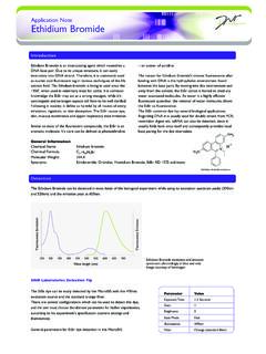
5 In contrast, detection of a phosphorylation-dependent epitope in a section of frozen tissue may require antigen retrieval and be dependent on amplified chromogenic visuali zation. This Guide provides information on the IHC/ICC variables listed below and is intended to assist in the design, optimization, and trouble shooting of IHC/ICC experiments. Examples of Fluorescence Detection by IHC and ICC. A. Fluorescence detection of V-Set and Ig Domain-Containing Protein 1 (VSIG1) in immersion-fi xed frozen sections of human testis using a Sheep Anti-Human VSIG1 Antigen Affi nity-Purifi ed Polyclonal Antibody (Catalog # AF4818). The tissue was stained using the NorthernLights 557-Conjugated Donkey Anti-Sheep IgG Secondary Antibody (Catalog # NL010; red) and counterstained with DAPI (blue). B. Fluorescence detection of Glial Fibrillary Acidic Protein (GFAP) expression in differentiating cultures of rat cortical stem cells using a Sheep Anti-Human GFAP Antigen Affi nity-Purifi ed Polyclonal Antibody (Catalog # AF2594).
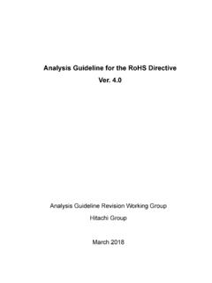
6 The cells were stained using the NorthernLights 557-Conjugated Donkey Anti-Sheep IgG Secondary Antibody (Catalog # NL010; red) and counter-stained with DAPI (blue).Examples of Chromogenic Detection by IHC and ICC. A. Chromogenic detection of Interferon Regulator Transcription Factor 8 (IRF8) expression in immersion-fi xed paraffi n-embedded sections of human lymphoma tissue using a Sheep Anti-Human IRF8 Antigen Affi nity-Purifi ed Polyclonal Antibody (Catalog # AF5117). The tissue was stained using the Anti-Sheep HRP-DAB Cell & Tissue Staining Kit (Catalog # CTS019; brown) and counter stained with hematoxylin (blue). B. Chromogenic detection of IFN- expression in human peripheral blood mononuclear cells that were stimulated with mg/mL of calcium ionomycin and 50 ng/mL of PMA using a Goat Anti-Human IFN- Antigen Affi nity-Purifi ed Polyclonal Antibody (Catalog # AF285).
7 The cells were stained using the Anti-Goat HRP-DAB Cell & Tissue Staining Kit (Catalog # CTS008; brown) and counterstained with hematoxylin (blue).VariableFactorsAntigenSpecies, expression levels, sample types, and subcellular locationEpitopeDependence on conformation or post-translation modifi cationSample TypeTissue or cellsSample PreparationTissue: embedded or frozenCells: adhered cell cultures or cell suspensionsAppropriate ControlsNo primary antibody, isotype control, absorption control, tissue type controlFixation MethodPerfusion or immersion (with or without freezing)Fixative*Formaldehyde, alcohols, or acetoneBlocking Reagent*Normal serum, bovine serum albumin (BSA), or non-fat milkAntigen Retrieval*Protease-Induced Epitope Retrieval (PIER) or Heat-Induced Epitope Retrieval (HIER) Detection MethodDirect or indirect (with or without amplifi cation)VariableFactorsPrimary Antibody*Monoclonal or polyclonalSecondary Antibody*Species and labelLabeling MethodFluorescence or chromogenicLabelFluorochromes and spectral propertiesChromogens.
8 3,3'-diaminobenzidine (DAB), 3-amino-9-ethylcarbazole (AEC) Counterstain*Fluorescence: 4,6'-diamidino-2-phenylindole (DAPI)Chromogenic: hematoxylinMounting ReagentFluorescence: anti-fade mounting mediumChromogenic: aqueous mounting mediumVisualization and AnalysisFluorescence microscope or standard light microscopeVariables for Experimental Design and to the IHC/ICC Method* These variables require optimization of additional factors including concentration, pH, temperature, incubation time, and diluent. This table is not intended to be comprehensive but represents themost commonly applied IHC/ICC methods at R&D Protocol Guide4 Tissue and cell samples must be appropriately harvested and prepared for each IHC/ICC study. To facilitate the required incubation steps, whole tissues must be cut into ultra thin (5 10 mm) sections or cut into smaller pieces for whole mount IHC. For ICC experiments, cells must be attached to a microscope slide or coverslip before commencing the staining procedure.
9 Sample preparation is also intimately linked to the method of fixation, which in turn is influenced by the desired detection technique (fluorescence versus chromo genic). In most circumstances, one experimental variable will determine the most appropriate method of sample preparation. For example, tissue that is immersion-fixed in formaldehyde must be paraffin-embedded and cut using a microtome. Alternatively, to detect a phosphorylation-dependent epitope, tissue may need to be snap frozen, which requires a cryostat for sample sectioning followed by alcohol TissueParaffin embedding offers the best option for long-term preservation of tissue samples. Tissue must be fixed before being embedded in paraffin. Fixation can be achieved by perfusion or immersion immediately following dissection, and typically requires 4 24 hours. It is not recommended to fix tissue for more than 24 hours because overfixation can cause masking of the antigen (see page 11).
10 If necessary, tissues can be transferred to alcohol after fixation until there is time for the embedding process. Because paraffin is immiscible with water, tissue must be dehydrated be fore adding molten paraffin wax. Dehydration is achieved by immersion in increasing concentrations of alcohol. This approach allows for a gradual change in hydropho bicity and minimizes cell damage. Following dehydration, the tissue is incubated with xylene to clear any remaining ethanol. Paraffin is typi cally heated to 60 C for embedding and is subsequently allowed to harden overnight. The tissue is subsequently cut with a sharp blade into ultra thin slices using a microtome. Sections are then dried onto micro scope slides and can be stored at room temperature for extended periods of time. Tissue sections must be rehydrated before commencing the IHC/ICC Protocol (see page 16). Paraffin is the cheapest and most commonly used substance for tissue embedding.