Transcription of Image Acquisition and Quality in Digital Radiography
1 53 RADIOLOGIC TECHNOLOGY, September/October 2016, Volume 88, Number 1 CEDirected ReadingThis article is a Directed Reading. Your access to Directed Reading quizzes for continuing education credit is determined by your membership status and CE preference. Shannon Alexander, MH A, (R)(T) Image Acquisition and Quality in Digital Radiography In 1895, German professors from the University of W rzburg per-formed experiments examining the properties of cathode rays and the effects of electrical current through low-pressure Continuing to investigate the effects of cathode rays passing through different types of vac-uum tube equipment, Professor Wilhelm Conrad Roentgen noticed that as the rays were emitted from the tube, a barium platinocyanide plate across the room would f Placing objects of differing thicknesses in the path of what he called x-rays, Roentgen was able to record varying transparencies of the objects on the photographic One object placed in the path of the x-ray was the hand of Roentgen s W hen the plate was developed, the shadows of his wife s hand bones were At that moment.
2 Wilhelm Conrad Roentgen became the first radiographer and the first roentgenogram was After weeks of intense and secretive experimentation, Roentgen reported his findings to the local medical soci-ety in Six years later, in 1901, he was awarded the Nobel Prize in Physics for his Producing X-RaysSince the first roentgenogram, advancements in medical imaging have been profound. Over the past 120 years, medical imaging has evolved from the invention of the x-ray tube by William Coolidge in 1915 to the subsequent development of imaging technologies, such as computed tomog-raphy and nuclear A lthough techniques and equipment have been refined over the past century, the production of x-rays essentially has remained the same. X-rays are created in a glass vacuum tube housed inside a protective metal After completing this article, the reader should be able to: Discuss the discovery of x-rays and how they are created.
3 Define Digital Radiography and describe how it differs from film-screen Radiography . Describe the Image Acquisition and readout process for computed Radiography , indirect Digital Radiography , and direct Digital Radiography systems. Identify various factors that affect Digital Image Quality and appearance. List factors used to determine radiation exposure to patients who have Digital Radiography examinations. Discuss changes that should occur in the workplace when implementing Digital Radiography . Medical imaging has undergone dramatic changes and technological breakthroughs since the introduction of Digital Radiography . This article presents information on the development of Digital Radiography and types of Digital Radiography systems. Aspects of Image Quality and radiation exposure control are highlighted as well. In addition, the article includes related workplace changes and medicolegal considerations in the Digital Radiography environment.
4 54 RADIOLOGIC TECHNOLOGY, September/October 2016, Volume 88, Number 1 CEDirected ReadingImage Acquisition and Quality in Digital Radiography capture exit beam rays was through conventional, or film-screen, In film-screen Radiography , the film acts as the medium for Image Acquisition , dis-play, and storage. Typically, a piece of radiographic film is placed on 1, or between 2, intensifying screens that emit light when struck by The light exposes the film in proportion to the amount and energy of the inci-dent x-rays on the The film is then processed with chemicals, and the Image appears on the sheet of A lthough film-screen Radiography can provide an adequate Image , problems can occur if the operator is not properly trained or makes small errors in technique. Poor Image Quality can result from incorrect deter-mination of radiation exposure settings. Fixed dose latitude and fixed nonlinear grayscale response limit the information that can be captured on the film and the potential for reducing patient In addition, film-screen Radiography can be expensive because of costs associated with purchasing film and processing materials.
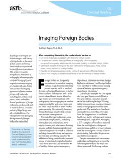
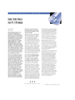
5 Storage, organization, retrieval, and dissemination of film also present expenses and obstacles for a radiology department, such as lost films or space for (see Figure 1).1 A negatively charged cath-ode sits at one end of the x-ray tube that also contains a tungsten The other end of the tube contains a positively charged anode and a thick copper rod with a small, angled tungsten W hen an x-ray is produced, the filament on the cathode end of the tube is heated, emitting electrons through the process of thermionic The electrons are accelerated by high voltage and focused to the anode W hen the electrons hit the tungsten target, x-rays are These rays are guid-ed by the angled tungsten target and directed toward the patient through the window of the tube The x-rays pass through the intended area of the patient s body where part of the x-ray beam is absorbed through On the opposite side of the body, detectors or film cap-tures the exit beam rays and a clinical Image is produced.
6 Image production and processing are dependent on the mechanism used to capture the attenuated Film-Screen RadiographyFollowing the discovery of the x-ray and the inven-tion of the radiograph, the most common way to Figure 1. The anatomy of the x-ray tube. ASRT anodeGlass enclosureWindowCathode assemblyTargetFocusing cupFilament 55 RADIOLOGIC TECHNOLOGY, September/October 2016, Volume 88, Number 1 CEDirected ReadingAlexanderbe altered to compensate for technique errors, best practice is to select the appropriate exposure technique factors for the patient s size and condition and follow the as low as reasonably achieveable (A LAR A) The final component of a Digital Radiography system is Image storage. Radiographic images must be stored for comparison and legal Shor t-ter m archiving generally is available on all systems, and long-term archival systems, such as PACS, are necessary to ensure the Image is available at a later date, sometimes years PACS is used for Digital storage of images and serves as a means to transfer images across networks and between medical Because images are stored and distributed electronically, many of the costs originally associated with storage and dissemination of film-screen radiographs are eliminated after transition-ing to Digital Radiography and ,6 Because Digital Radiography practice still is transition-ing to completely Digital technology, many terms and techniques relating to DR are not yet standard or univer-sally This article focuses on 3 types of Digital Radiography .
7 Computed Radiography , indirect Digital radi-ography, and direct Digital Radiography . Computed Radiography Computed Radiography (CR) was introduced commercially in 1983 and initially was referred to as cassette-based Digital Although cassetteless CR systems are available, only cassette-based CR sys-tems will be discussed. Acquiring an Image using CR is comparable to acquiring an Image using film-screen The radiographer positions the patient, centers the x-ray beam, and sets technical details. The primary difference between film-screen Radiography and CR is the composition of the The CR cassette, or Image plate, comprises various layers needed to acquire an Behind the protec-tive layer of the cassette is the phosphor layer. This layer is known as the active layer because it contains a photo-stimulable phosphor that collects electrons during an exposure. The next layer is electroconductive to absorb and reduce static electricity and generally is made of felt or foam to prevent dust accumulation.
8 Next, a support layer gives the imaging plate some strength. Newer cassettes might contain a light shield or color layer that absorbs Digital RadiographyDigital imaging has been defined as any imaging Acquisition process that produces an electronic Image that can be viewed or manipulated on a computer. 5 This definition applies to many types of images, but Digital images acquired using radiation are referred to as Digital A lthough Digital subtraction angi-ography (a type of f luoroscopy used in interventional radiology), developed in 1977, is thought to be the first Digital imaging system, the first Digital system for gen-eral Radiography was introduced in 1980 and required cassette-based storage-phosphor Image Over the next decade, many improvements were made and its use became By 1990, technology had evolved, and a new type of Digital Radiography system appeared that used sensors to detect attenuated x-rays and did not require cassettes.
9 These completely Digital systems facilitated the use of PACS to store and trans-fer Although film-screen and cassette-based systems still are used by some providers, the trend is moving toward completely Digital systems. In 2013 it was estimated that newly installed film-screen systems made up only 1% of the In 2016, the American Registry of Radiologic Technologists (AR RT) Board of Trustees voted to remove film-screen topics from the Image production section of the certification exam and expand Digital imaging topics effective January 1, 2 In addition, beginning in 2017, Medicare is going to reduce payments by 20% for providers using film-screen systems and 7% to 10% for providers using cassette-based systems, further solidifying the adoption of completely Digital Radiography physical principles of creating x-rays with Digital Radiography are the same as with film-screen Radiography .
10 However, data Acquisition , Image process-ing, viewing, and storage In place of film and intensifying screens, Digital Radiography uses detectors to measure and convert the attenuated rays from the patient into electronic These signals are then recorded, digitized, and quantified into a grayscale that represents the amount of x-ray energy that was depos-ited at a given Postprocessing software organizes the data into a clinically meaningful Image that is displayed on a computer A lthough the Image displayed can 56 RADIOLOGIC TECHNOLOGY, September/October 2016, Volume 88, Number 1 CEDirected ReadingImage Acquisition and Quality in Digital Radiography After exposure, the radiographer attaches patient demographic information to the cassette via a barcod-ing system and then inserts the cassette into a reader to obtain the The imaging plate is removed from the cassette and scanned by a Typically, manufac-turers use a helium laser or solid-state laser diodes for the signal extraction The laser scans the imag-ing plate, producing a photostimulated luminescence, which is converted into an electrical signal and digitized by an analog-to- Digital digitization, a computer processes the signal and displays the Digital Image on a At this stage, Image Quality can be assessed and postprocessing adjustments can be made before images are sent to the radiologist for Typically, the entire stimulating light but ref lects emitted Last.