Transcription of Abdominal Aortic Aneurysms - American Society of ...
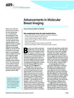
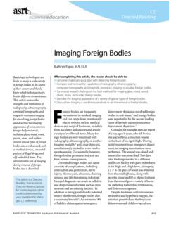
1 145 RADIOLOGIC TECHNOLOGY, November/December 2016, Volume 88, Number 2 CEDirected ReadingThis article is a Directed Reading. Your access to Directed Reading quizzes for continuing education credit is determined by your membership status and CE preference. The aorta is the largest artery in the human body. Originating from the left ventricle, it extends into the chest and down into the abdomen. The vessel is the main conduit for distributing oxy-genated blood from the heart to the body. Abdominal Aortic aneurysm (A A A) is a localized enlargement with-in the Abdominal cavity. Anatomy and PhysiologyThe aorta is the major artery of the body carrying oxygenated blood from the heart through the thoracic and Abdominal cavities of the body (s e e Figure 1).
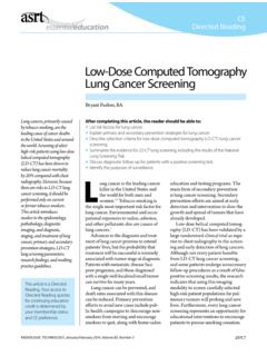
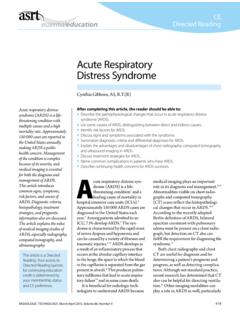
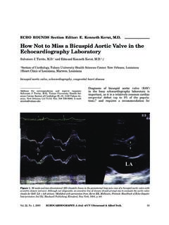
2 The aorta attaches superiorly to the left ventricle. Blood is pumped through the aorta with every left ventricular contraction. A 3-leaf let valve at the junction of the left ventricle and aorta ( Aortic valve) prevents retro-grade f low of blood into the aorta is divided into 4 sec-tions: ascending aorta , Aortic arch, thoracic aorta , and Abdominal aorta . The ascending aorta , which rises superiorly from the left ventricle, is approximately 3 cm in diameter but can vary depending on patient sex, age, and size. At the root of the ascending aorta are 3 anatomical dilatations called the Aortic sinuses. From the left and right Aortic sinuses arise the left and right coronary arteries, respectively. The coronary arteries vascularize the heart (s e e Figure 2).
3 The posterior sinus has no coronary completing this article, the reader should be able to: Describe Aortic anatomy. State the preventive screening recommendations for Abdominal Aortic aneurysm (AAA). Identify the most common locations of AAA occurrence. Recall the preferred imaging modalities for AAA and their specific applications. Discuss treatments for Aortic aneurysm (A A A) is a significant disease affecting the circulatory system. Risk factors include smoking , hypertension, sex, and a possible hereditary predisposition. A A As remain asymptomatic for years, and various imaging methods are used in their detection, diagnosis, and treatment. This article reviews the anatomy and physiology of the aorta as well as the signs and symptoms, pathophysiology, epidemiology, and risk factors for the development of A A A.
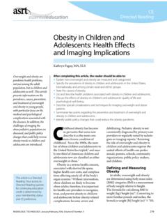
4 The use of ultrasonography and other imaging modalities for pre- and post-treatment is discussed, as is endovascular Aortic S Legg, PhD, (R)(CT)(QM), FASRTLynn M Legg, MBA, (R)(M) Abdominal Aortic AneurysmsIliacInferior mesentericRenalBrachiocephalicCeliacAdre nalAortaSubclavianLeft carotidSuperior mesentericFigure 1. Pathway of the aorta from the heart to the iliac artery bifurcation. ASRT TECHNOLOGY, November/December 2016, Volume 88, Number 2 CEDirected ReadingAbdominal Aortic AneurysmsThe aorta enters the Abdominal cavity through the diaphragm and then divides into 2 major vessels: common iliac arteries and the median sacral artery. Important branches of the Abdominal aorta are the lumbar and musculophrenic arteries, renal and suprare-nal arteries, and arteries that vascularize the Abdominal viscera, such as the celiac trunk and the superior and inferior mesenteric , all arteries have 3 layers.
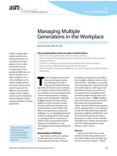
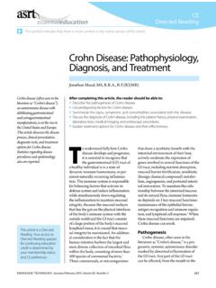
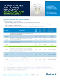
5 The layers of the aorta are composed of smooth muscle, nerves, intimal cells, and extracellular matrix. The Aortic wall is divided into 3 layers (from external to lumen): tunica externa (or tunica adventitia), tunica media, and tunica intima (see Figure 3). The vascular supply to the tunica externa and tunica media is pro-vided by an extensive network of small blood vessels known as the vasa f lows through the aorta in pulses due to contraction of the left ventricle. The elasticity of the aorta allows it to expand and contract as the heart pumps blood through it. The tunica media contains collagen and elastin filaments that allow it to stretch and contract in response to the pulsatile f low of blood, assisting in the propulsion of blood through the circulatory system.
6 For As the aorta rises superiorly from the heart, it makes a downward curve, which is known as the Aortic arch. The Aortic arch often is visible on chest radiographs; from it arises arteries supplying blood to the head, neck, and upper limbs the brachiocephalic artery (which bifurcates to form the right subclavian artery and right common carotid artery), left common carotid artery, and left subclavian artery. The Aortic arch also has function in homeostasis, containing barorecep-tors and chemoreceptors that send information about carbon dioxide levels and blood pH to the medulla ,3 Beyond the Aortic arch, the aorta descends caudally, which is called the descending aorta . The descending aorta is divided into 2 parts based on location. The initial section of the aorta that passes through the medi-astinum and chest is called the thoracic aorta .
7 Many branches originate from the thoracic descending aorta , including intercostal and subcostal arteries, bronchial arteries, and branches that supply blood to the esopha-gus, mediastinum, pericardium, and 2. Aortic arch and its associated arteries. ASRT 3. Layers of the Aortic wall. ASRT externaTunica mediaEndotheliumTunica intimaBasement membraneBrachiocephalic arteryAscending aortaRight coronary arteryLeft coronary arteryLeft common carotid arteryLeft subclavian arteryAortic archDecending aorta (thoracic) Aortic sinusesarteryy 147 RADIOLOGIC TECHNOLOGY, November/December 2016, Volume 88, Number 2 CEDirected ReadingLegg, LeggPrevalenceA A As are the most common arterial aneurysm, with approximately 80% occurring below the renal arteries at the area of Aortic Studies of adults 50 years and older indicate an A A A prevalence of to in men and to in ,10 Worldwide, the prevalence is higher in western countries than in Asiatic countries, and higher in Australia than in the United States and If diagnosed before becoming symp-tomatic, an A A A can be treated and cured.
8 However, a ruptured A A A is considered a medical emergency, with a 59% to 90% mortality rate if it occurs outside a hospital ,12 The operative mortality for A A A, defined as death regardless of cause occurring within 30 days after surgery,13 is approximately 40%.9 Mortality rates for elective (ie, nonemergency) A A A repair are significantly lower, ranging from to -16 Therefore, careful screening to detect A A As and followup when diagnosed are necessary, specifically with at-risk diameter of the aneurysm inf luences the poten-tial for rupture. For A A As cm to cm in diameter, the annual risk for rupture is negligible. The risk is 1% for Aneurysms that are between cm and cm, and example, as the left ventricle contracts (systole), blood is forced into the aorta , which expands.
9 This distention provides the potential energy that helps to main-tain blood pressure during ,3 The aorta s elastic recoil also helps the heart conserve energy. The pressure of the blood through the aorta , termed the pulse pressure, equals systolic pressure minus diastolic measure of the pressure of blood f low through the aorta is called mean arterial pressure (M AP), often abbreviated as Pmean. M AP is a measure of the cardiac output, systemic vascular resistance, and central venous pressure. M AP is highest at the aorta and decreases across the cir-culatory system as the aorta branches into arteries and then into arterioles, capillar-ies, and the velocity of the blood pulse wave also aids in assessing Aortic function. Pulse wave velocity (PW V) is a measure of Aortic stiffness and can be measured invasively via f low meters or catheter-based pressure probes in patients undergoing cardiac catheterization,4,5 or noninvasively with The aorta stiffens with age, which results in increased5: Blood pressure.
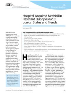
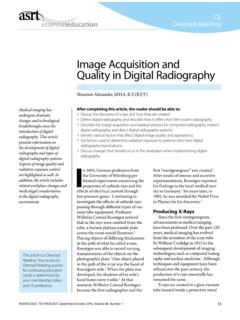
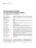
10 R isk of developing blood pressure-related diseases and pathologies. Correlation with cardiovascular events and mor-ta l it y. PathophysiologyIn general, an A A A is a focal, localized enlarge-ment (dilatation) of a section of the Abdominal aorta of cm or greater, or when the diameter of the dilatation is 50% larger than normal (see Figure 4).7 Dilatation of the Aortic wall occurs when the tunica media weakens. The pulsating force of the blood through the aorta causes the tunica intima and externa to stretch and expand. As the vessel wall layers progres-sively weaken, the aneurysm continues to enlarge, often resulting in a fatal rupture. Figure 4. Diagram of an Abdominal Aortic aneurysm (A A A) as compared to a normal aorta . ASRT 2016. Normal aortaAbdominal Aortic aneurysm (infrarenal)148 RADIOLOGIC TECHNOLOGY, November/December 2016, Volume 88, Number 2 CEDirected ReadingAbdominal Aortic Aneurysmsundetected for years.