Transcription of Introduction to Medical Image Processing
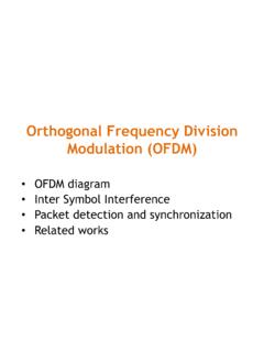
1 1 Introduction to Medical Image Processing Essential environments of a Medical imaging system Image Processing may be a post-imaging or pre-analysis operator. Functions of Image Processing and Image analysis may overlap each other. Imaging System System Image Processing Images Feature Images Energy Image Analysis Subject 2 Goals of Medical Image analysis techniques: Quantification: Measuring the features on Medical images, eg., helpng radiologist obtain measurements from Medical images ( , area or volume). * To make the features measurable, it is necessary to extract objects from images by segmentation.
2 Computer Aided Diagnosis (CAD): given measurements and features make a diagnosis. Help radiologists on their diagnosis procedure for accuracy and efficiency. Evaluation and validation techniques. 3 General Image analysis (regardless of its application) encompasses: incorporation of prior knowledge classification of features matching of model to sub-images description of shape many other problems and approaches of About digital Image Processing What is an Image ? Formal definition: A digital Image is a multi-dimensional signal that is sampled in space and/or time and quantized in amplitude.
3 Looser definition: An Image is a picture." The brightness values in the picture may represent distance, reflectivity, density, temperature, etc. The Image may be 2-D (planar), 3-D (volumetric), or N-D. 4 Image elements: An Image may be composed of: 2-D: pixels = picture elements 3-D: voxels = volume elements 4-D and higher: hypervoxels = hypervolume elements What do images represent? For Medical images, pixels may represent parameters such as: X-ray attenuation (density) Water (proton) density Acoustic impedance (distribution) Optical reflectivity (or impedance) Electrical activity , etc.
4 How are images processed? Manual analysis: processed by human Semi-automatic analysis: human and computer work together to process Image Automatic analysis: computer processes Image , human reviews the results 5 Purposes of Image Processing : Preprocess Image to reduce noise and blur (filtering) Identify structures within the Image (segmentation) Extract useful information from the Image (quantification) Prepare the Image for visualization (enhancement, reconstruction) * Exact Processing steps depend on the application. What we do to images: Enhancement: Noise reduction Deblur Improve contrast Identify structures in Image (segmentation): Identify homogeneous regions in an Image (label Image pixels = segmentation) Measure: 6 For example: Heart chamber volumes , Heart wall motion , Brain activity, Fetus size/gender , Lesion size and extent.
5 Visualize: For example: Surgical/Therapy planning , Image -guided surgery Processing verse Analysis Medical Image Processing Deals with the development of problem specific approaches to enhance the raw Medical data for the purposes of selective visualisation as well as further analysis. Medical Image analysis Concentrates on the development of techniques to supplement the usually qualitative and frequently subjective assessment of Medical images by human experts. Provides quantitative, objective and reproducible information extracted from the Medical images 7 So many subjects: 57 chapters in Handbook of Medical Image Processing and Analysis , by Isaac Bankman (Ed.)
6 , Academic Press, 2009. I Enhancement; Fundamental Enhancement Techniques; Adaptive Image Filtering; Enhancement by Multiscale Nonlinear Operators; Medical Image Enhancement with Hybrid Filters; II Segmentation; Overview and Fundamentals of Medical Image Segmentation; Image Segmentation by Fuzzy Clustering: Methods and Issues; Segmentation with Neural Networks; Deformable Models; Shape Information in Deformable Models; Gradient Vector Flow Deformable Models; Fully Automated Hybrid Segmentation of the Brain; Unsupervised Tissue Classification; Partial Volume Segmentation with Voxel Histograms; Higher Order Statistics for Tissue Segmentation; III Quantification; Two-dimensional Shape and Texture Quantification; Texture Analysis in Three Dimensions for Tissue Characterization; Computational Neuroanatomy Using Shape Transformations; Tumor Growth Modeling in Oncological Image Analysis; Arterial Tree Morphometry; Image -Based Computational Biomechanics of the Musculoskeletal System; Three-Dimensional Bone Angle Quantification; Database Selection and Feature Extraction for Neural Networks.
7 Quantitative Image Analysis for Estimation of Breast Cancer Risk; Classification of Breast Lesions in Mammograms; Quantitative Analysis of Cardiac Function; Image Processing and Analysis in Tagged Cardiac MRI; Analysis of Cell Nuclear Features in Fluorescence Microscopy Images; Image Interpolation and Resampling; IV Registration; Physical Basis of Spatial Distortions in Magnetic Resonance Images; Physical and Biological Bases of Spatial Distortions in PET Images; Biological Underpinnings of Anatomic Consistency and Variability in the Human Brain; Spatial Transformation Models; Validation of Registration Accuracy; Landmark-based Registration Using Features Identified through DifferentialGeometry; Image Registration Using Chamfer Matching; Within-Modality Registration Using Intensity-Based Cost Functions; Across-Modality Registration Using Intensity-Based Cost Functions; Talairach Space as a Tool for Intersubject Standardization in the Brain; Warping Strategies for Intersubject Registration; Optimizing the Resampling of Registered Images; Clinical Applications of Image Registration.
8 Registration for Image -Guided Surgery; Image Registration and the Construction of Multidimensional Brain Atlases; V Visualization; Visualization Pathways in Biomedicine; Three-Dimensional Visualization in Medicine and Biology; Volume Visualization in Medicine; Fast Isosurface Extraction Methods for Large Image Data Sets; Computer Processing Methods for Virtual Endoscopy; VI Compression, Storage, and Communication; Fundamentals and Standards of Compression and Communication; Medical Image Archive and Retrieval; Image Standardization in PACS; Imaging and Communication in Medical and Public Health Informatics; Dynamic Mammogram Retrieval from Web-Based Image Libraries; Quality Evaluation for Compressed Medical Images: Fundamentals; Quality Evaluation for Compressed Medical Images: Diagnostic Accuracy; Quality Evaluation for Compressed Medical Images: Statistical Issues; Three-Dimensional Image Compression with Wavelet Transforms.
9 8 Why Image Enhancement? Can t distinguish between tissues The nature of the physiological system under investigation and the procedures used in imaging may diminish the contrast and the visibility of details. Data is too noisy for computer algorithm to perform well Medical images are often deteriorated by noise due to various sources of interference and other phenomena that affect the measurement processes in imaging and data acquisition systems. Imaging artifacts interfere with visualization or computer Processing How to Enhance Image ? By operations to Increase contrast Remove noise Emphasize edges: Edge boost, Unsharp masking.
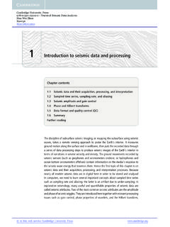
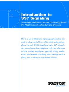
10 Modify shapes 9 * Image enhancement techniques range from linear to nonlinear, from fixed to adaptive, and from pixel-based to multiscale methods, .. Contrast Enhancement by Histogram Equalization 10 Enhancement by adaptive wavelet shrinkage denoising (only subimages with diagnostic information are reconstructed). 11 Enhancement by adaptive filtering: noise or speckle reduction. Adaptive Image filtering needs some a priori information about the Image . ( This is an Image modeling problem) Geometric or statistical information are the primary information used. 12 Medical Image Segmentation Segmentation, separation of structures of interest from the background and from each other, is an essential analysis function for which numerous algorithms have been developed in the field of Image Processing .