Transcription of Introduction to Small-Angle X-ray Scattering
1 Introduction to Small-Angle X-ray Scattering Thomas M. Weiss Stanford University, SSRL/SLAC, BioSAXS beamline BL 4-2 BioSAXS Workshop, March 28-30, 2016. Sizes and Techniques Diffraction and Scattering Scattering of X-rays from a single electron Ie Thomson formula for the scattered intensity from a single electron 2 . I0. 1 cos( 2 ) 1. I e r0. 2. 2. I0. 2 r 2. e 15. r0 2. 2 . 817 10 m I 0 : Intensity of incoming X - rays mc 2 : angle of observatio n I e : Intensity of scattered X - rays Classical electron radius The Thomson formula plays a central role for all Scattering calculations involving absolute intensities.
2 Typically calculated intensities of a given sample will be expressed in terms of the Scattering of an isolated electron substituted for the sample. In small angle Scattering the slight angle dependence (the so-called polarization factor). in the Thomson formula can be neglected. Interference of waves waves have and amplitude and phase interference leads to fringe pattern ( water waves). the fringe pattern contains the information on the position of the sources ( structure). in X-ray diffraction the intensities (not the amplitudes) of the fringes are measured phase problem.
3 Constructive destructive Scattering from two (and more) electrons Scattering vector q k k 0. two electrons 2 4 sin . with k s and q q 2. F (q ) f e exp( i q r i ) f e (1 exp( i q r )). i 1. Note: generalized to N electrons F(q) is the Fourier N Transform of the F (q ) f e exp( i q r i ) spatial distribution i 1 of the electrons averaged over all orientations N. sin( qr ). F (q ) fe qr i 1. using the continuous (radial) distribution ( r ) of the electron cloud in an atom . sin( qr ). dr ( r ) r f (0 ) Z. F (q ) f (q ) with 2. 0. qr atomic Scattering factor Scattering from Molecules The Scattering amplitude or form factor, F(q), of an isolated molecule with N atoms can be determined in an analogous manner: N.
4 The Fourier Transform F (q ) f i ( q ) exp( i q r i ). of the atomic distribution i 1. The scattered intensity from the isolated molecule is then N N.. 2. I (q ) F (q ) f i ( q ) f j ( q ) exp( i q ( r i r j )). i 1 j 1. In solution: N N. average over all orientations sin( qr ij ). sin( qr ij ). I (q ) fi (q ) f j (q ). qr ij due to solution average only exp( i q ( r i r j )) i 1 j 1. qr ij interatomic distances are Debye formula measured, not atomic coordinates Scattering from Molecules N N. sin( qr ij ). I (q ) fi (q ) f j (q ).
5 Qr ij i 1 j 1. Each atomic distance rij in the molecule adds a sinx/x like term to the Scattering intensity small distance low frequency in sinx/x dominate signal at high q large distance high frequencies in sinx/x dominate the signal at low q Scattering Intensity . I ( q ) F (q ) F (q ) FT [ ( r )] FT [ ( r )] FT [ ( r ) ( r )]. (r ). Autocorrelation function The measured Scattering intensity is the spherically averaged Fourier transform of the auocorrelation of the electron density of the particle Autocorrelation (r ) (r ) ( r ) ( r u ) ( r ) dV u Vu Autocorrelation (r ) (r ) ( r ) ( r u ) ( r ) dV u Vu Autocorrelation (r ) (r ) ( r ) ( r u ) ( r ) dV u Vu Autocorrelation (r ) (r ) ( r ) ( r u ) ( r ) dV u Vu Autocorrelation (r ) (r ) ( r ) ( r u ) ( r ) dV u Vu Autocorrelation (r ) (r ) ( r ) ( r u ) ( r )
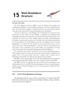
6 DV u Vu For a homogeneous particle Spherical average Characteristic Function (r ) r V ( r ) (r ) 0 (r ) (r ) / (0 ). with ( 0 ) V. 2. 0 r V. probability of finding a point Pair distance distribution function: within the particle at a distance r from a given point . p ( r ) r ( r ) Vr 0 ( r ). 2 2 2. Pair distance distribution function p(r). The p(r) function represents the histogram of distances between pairs of points within the particle. Dmax is the maximum diameter in the particle. Measured Scattering intensity Pair distance distribution.
7 Dmax p ( r ) 4 I ( q ) qr sin( qr ) dq 0. Scattering from model structures Adopted from Svergun & Koch, SAS studies of biological macromolecules in solution , Rep. Prog. Phys. 66 (2003) 1735-1782, Fig. 5 (c)I. Particles in Solution For solution Scattering we typically require the following characteristics: Monodisperse, identical particles i j ( q ) i1 ( q ) j Uncorrelated, no inter-molecular interactions present I (q ) n j i j (q ). j I ( q ) Ni 1 ( q ). Background Scattering and X-ray Contrast I solution ( q ) I solvent ( q ) I particle ( q ).
8 The solvent Scattering background must be properly subtracted to obtain the signal from the particles the contrast, that makes the particles visible for X-rays, is the difference in electron density of the particle versus the solvent ( (r ) s ). 2. Protein solution Scattering data weak level of Scattering at small angles drops off quickly for higher angles due to low contrast Scattering level of background and sample is very similar except for the lowest angles background and sample Scattering need to be measured with high accuracy a 1mg/ml solution of a globular protein of the size of lysozyme (14kD) scatters on the order of: 1 out of 106 incident photons.
9 One in a million! . X-ray Contrast and Contrast Variation change contrast by adding salts ( Substance Average Contrast CsBr), sucrose or glycerol to the solvent (x1010 cm-2) but that changes the chemical Protein environment for the particles other possibility to change contrast is Nucleic Acid anomalous Scattering Fatty Acid Carbohydrates Note: Contrast variation is widely used in neutron Scattering , due to the large Scattering length difference of hydrogen and deuterium Introducing the Radius of Gyration Rg2 is the average electron density r (r ) d r 2.
10 Weighted squared distance of the 2. scatters from the centre of the Rg . object (r ) d r 2. 1 Solid sphere radius R: 1 3. 12 Rg = (3/5) R. Thin rod length L. Rg2 =(12+ 12+ 12+ 22+ 22+ 32 )/6=20/6. Rg = (1/12) L. Rg= = Thin disk radius R: Rg = (1/2) R. The Guinier approximation The low-q region of the Scattering curve is characteristic for the overall dimension of the particle. 1. Guinier region I(q) 2. lim I I 0 exp( . 2. q Rg ). q 0 3. I0 is proportional to Mw Radius of gyration: size of the particle q The Guinier Plot . Plot ln I against q2 Straight line, slope Rg/3.