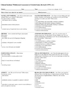
Transcription of Low QRS voltage and its causes - UMEM
1 Available online at Journal of Electrocardiology 41 (2008) 498 500. Low QRS voltage and its causes John E. Madias, MD . Mount Sinai School of Medicine of the New York University, New York, NY, USA. Division of Cardiology, Elmhurst Hospital Center, Elmhurst, NY, USA. Received 11 April 2008; revised 23 June 2008; accepted 23 June 2008. Abstract Electrocardiographic low QRS voltage (LQRSV) has many causes , which can be differentiated into those due to the heart's generated potentials (cardiac) and those due to influences of the passive body volume conductor (extracardiac). Peripheral edema of any conceivable etiology induces reversible LQRSV, reduces the amplitude of the P waves and T waves, decreases the duration of P waves, QRS.
2 Complexes, and QT intervals, and alters in turn the measurements of the signal-averaged electro- cardiogram and T wave alternans, all with enormous clinical implications. 2008 Elsevier Inc. All rights reserved. Keywords: ECG; Low QRS voltage ; causes of low QRS voltage ; Passive body volume conductor; Electrical resistivity of body tissues Low electrocardiographic QRS voltage (LQRSV) is pericardium is part of the heart, or the extracardiac causes traditionally defined by zenith-to-nadir QRS amplitudes of because the site of generation of the cardiac potentials are the QRS complexes of less than mV in all the frontal inside the locus of pericardial fluid accumulation.
3 Occasion- leads and less than mV in all the precordial ally, both cardiac and extracardiac causes of LQRSV are However, the remarks that follow pertain both to electro- simultaneously in operation in the same patient. cardiograms (ECGs) with LQRSV and to any attenuation of the QRS voltage based on the comparison of at least 2 ECGs, Cardiac causes of LQRSV. even if none of them satisfy the above-cited criteria for LQRSV. Occasionally, LQRSV in the ECG may not have an Multiple myocardial infarctions may lead to LQRSV. apparent explanation; and thus, in normal subjects, it is because of cancellations and diminished electromotive force considered a normal variant.
4 Low ECG QRS voltage may be generation; LQRSVand QRS notches are seen in conjunction noted only in the limb leads (a frequent encounter), the with severe post myocardial infarction Infiltra- precordial leads, or both. Low ECG QRS voltage in limb tive cardiomyopathies, a prototypical example being amy- leads with normal QRS precordial amplitudes,2 or LQRSV in loidosis, may lead to LQRSV involving both the limb and the limb leads with high QRS complexes in the precordial leads precordial leads,5 which occurs despite the marked cardiac with poor R-wave progression ( Goldberger triad )3 have hypertrophy or dilatation. Other infiltrative cardiomyopathies been described in patients with dilated cardiomyopathy.
5 Are reputed to be associated with LQRSV, but literature There are many causes of LQRSV, and they can be review does not provide relevant information. Myocarditis is differentiated into those due to the deficient heart's generated associated with LQRSV attributed to the ailing myocytes,6. potentials (cardiac causes ) and those due to the attenuating although extracardiac influences may also contribute to the influences of the pericardial space and pericardium, or the LQRSV. 7 Reduction of QRS voltage (not necessarily passive body volume conductor, enveloping the heart LQRSV) follows reduction of cardiac volumes due to various (extracardiac causes ).
6 Pericardial pathology, particularly pathologies, hemorrhage, or hypovolemia ( Brody effect ).8. pericardial effusion, can be categorized among the cardiac This is probably the mechanism for the LQRSV in patients causes of LQRSV, considering that the visceral and parietal with Addison's disease, although pulmonary congestion and/. or peripheral edema (PERED) may contribute to LQRSV. Division of Cardiology, Elmhurst Hospital Center, 79-01 Broadway, (vide infra).9 Extensive skin burns may lead to hypovolemia Elmhurst, NY 11373, USA. Tel.: +1 718 334 5005; fax: +1 718 334 5990. and cause LQRSV, although associated hypoalbuminemia E-mail address: may contribute (vide infra).
7 An increased hematocrit leads to 0022-0736/$ see front matter 2008 Elsevier Inc. All rights reserved. Madias / Journal of Electrocardiology 41 (2008) 498 500 499. 25 26. LQRSV because of the reduction in disparity of electrical pneumopericardium, pneumomediastinum, and pneu- resistivity of the intracardiac blood mass and the surrounding mothorax, particularly left sided,27 are associated with myocardium, which attenuates the radial depolarization LQRSV. Pulmonary edema 28 and bronchopulmonary forces, as per the Brody ,10 Intracardiac injection of lavage 29 result in LQRSV because of decreased lung contrast media leads to some attenuated QRS voltage11; impedance by way of increased water content.
8 Pneumonia, however, the authors attributed this to 3-vessel coronary with extensive pulmonary infiltrates, and adult respiratory artery disease. The voluminous literature on the topic focuses distress syndrome ( wet lung ) are expected to lead to on QTc and other repolarization This author similar ECG findings, although nothing to this effect has suspects that the LQRSV may be due to increased resistivity been described heretofore. Pleural effusion, particularly left of the contrast media,13 inducing the Brody effect; and it must sided and in the absence of congestive heart failure, causes be different for ionic, nonionic, hyperosmolar, isoosmolar, LQRSV with an inverse relation between the extent of the and hypoosmolar contrast media.
9 Although LQRSV is rarely effusion and the amplitude of QRS Extensive found in hypothyroidism in the absence of pericardial subcutaneous emphysema, with retropneumoperitoneum and effusion, it has been suggested that hypothyroidism per se mediastinal emphysema, is associated with LQRSV. 31. contributes to Change in the position of the heart and its relationship with the chest wall, and certain ECG leads32 must be influential in LQRSV in some of the above-discussed Pericardial causes of LQRSV conditions. Obesity is linked to reversible In the past few years, it has been observed that PERED of Pericardial effusion leads to LQRSV, the mechanism any conceivable etiology is predictably linked to purported to be that of a short-circuiting of the heart's Thus, PERED seen in association with sepsis,34 certain drugs potentials as they are transmitted to the body surface.
10 (eg, nonsteroidal anti-inflammatory drugs35 and the antidia- however, the mechanism may be more complex15 and may betic thiazolidinediones [unpublished data]), cor include even the intrapericardial pressure, like in tamponade, pulmonale,34 perioperative fluid load administration, even as the primary reason, along with the ,17 The in the presence of normal left ventricular function,36 chronic delays in recovery of LQRSV after pericardiocentesis or renal failure, particularly during the predialytic state,37. alleviation of tamponade suggest that the effects on the ECG. congestive heart failure,38 hepatic cirrhosis,39 and numerous in pericarditis/pericardial effusion/tamponade are multifac- other conditions result in reversible LQRSV.